2W0F | pdb_00002w0f

Potassium Channel KcsA-Fab Complex with Tetraoctylammonium


Experimental Data Snapshot

  • Method: X-RAY DIFFRACTION
  • Resolution: 2.40 Å
  • R-Value Free: 
    0.241 (Depositor), 0.240 (DCC) 
  • R-Value Work: 
    0.200 (Depositor), 0.198 (DCC) 
  • R-Value Observed: 
    0.204 (Depositor) 

Starting Model: experimental
View more details

wwPDB Validation 3D Report Full Report

Validation slider image for 2W0F

Ligand Structure Quality Assessment 


This is version 1.4 of the entry. See complete history

Literature

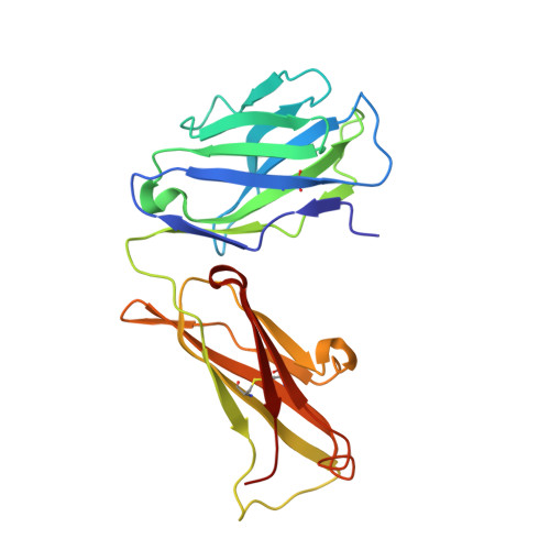
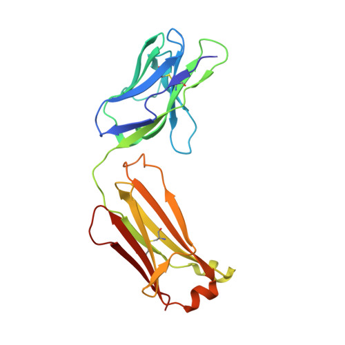
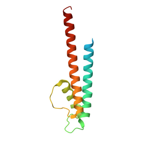
Structures of Kcsa in Complex with Symmetrical Quaternary Ammonium Compounds Reveal a Hydrophobic Binding Site.

Lenaeus, M.J.Burdette, D.Wagner, T.Focia, P.J.Gross, A.

(2014) Biochemistry 53: 5365

  • DOI: https://doi.org/10.1021/bi500525s
  • Primary Citation Related Structures: 
    2JK5, 2W0F, 4UUJ

  • PubMed Abstract: 

    Potassium channels allow for the passive movement of potassium ions across the cell membrane and are instrumental in controlling the membrane potential in all cell types. Quaternary ammonium (QA) compounds block potassium channels and have long been used to study the functional and structural properties of these channels. Here we describe the interaction between three symmetrical hydrophobic QAs and the prokaryotic potassium channel KcsA. The structures demonstrate the presence of a hydrophobic pocket between the inner helices of KcsA and provide insight into the binding site and blocking mechanism of hydrophobic QAs. The structures also reveal a structurally hidden pathway between the central cavity and the outside membrane environment reminiscent of the lateral fenestration observed in sodium channels that can be accessed through small conformational changes in the pore wall. We propose that the hydrophobic binding pocket stabilizes the alkyl chains of long-chain QA molecules and may play a key role in hydrophobic drug binding in general.


  • Organizational Affiliation
    • Department of Molecular Pharmacology and Biological Chemistry, Northwestern University Medical School , 303 East Chicago Avenue, Chicago, Illinois 60611, United States.

Macromolecule Content 

  • Total Structure Weight: 61.65 kDa 
  • Atom Count: 4,407 
  • Modeled Residue Count: 532 
  • Deposited Residue Count: 555 
  • Unique protein chains: 3

Macromolecules

Find similar proteins by:|  3D Structure
Entity ID: 1
MoleculeChains  Sequence LengthOrganismDetailsImage
ANTIBODY FAB FRAGMENT LIGHT CHAIN219Mus musculusMutation(s): 0 
Find similar proteins by:|  3D Structure
Entity ID: 2
MoleculeChains  Sequence LengthOrganismDetailsImage
ANTIBODY FAB FRAGMENT HEAVY CHAIN212Mus musculusMutation(s): 0 
Find similar proteins by:|  3D Structure
Entity ID: 3
MoleculeChains  Sequence LengthOrganismDetailsImage
VOLTAGE-GATED POTASSIUM CHANNEL124Streptomyces lividansMutation(s): 0 
Membrane Entity: Yes 
UniProt
Find proteins for P0A334 (Streptomyces lividans)
Explore P0A334 
Go to UniProtKB:  P0A334
Entity Groups
Sequence Clusters30% Identity50% Identity70% Identity90% Identity95% Identity100% Identity
UniProt GroupP0A334
Sequence Annotations
Expand
Reference Sequence

Small Molecules

Ligands 5 Unique
IDChains Name / Formula / InChI Key2D Diagram3D Interactions
DGA

Query on DGA


Download:Ideal Coordinates CCD File
F [auth C]DIACYL GLYCEROL
C39 H76 O5
UHUSDOQQWJGJQS-QNGWXLTQSA-N
HX0

Query on HX0


Download:Ideal Coordinates CCD File
G [auth C]N,N,N-trioctyloctan-1-aminium
C32 H68 N
CHYBTAZWINMGHA-UHFFFAOYSA-N
F09

Query on F09


Download:Ideal Coordinates CCD File
E [auth C]NONAN-1-OL
C9 H20 O
ZWRUINPWMLAQRD-UHFFFAOYSA-N
CO

Query on CO


Download:Ideal Coordinates CCD File
D [auth C]COBALT (II) ION
Co
XLJKHNWPARRRJB-UHFFFAOYSA-N
K

Query on K


Download:Ideal Coordinates CCD File
H [auth C]
I [auth C]
J [auth C]
K [auth C]
L [auth C]
H [auth C],
I [auth C],
J [auth C],
K [auth C],
L [auth C],
M [auth C],
N [auth C]
POTASSIUM ION
K
NPYPAHLBTDXSSS-UHFFFAOYSA-N

Experimental Data & Validation

Experimental Data

  • Method: X-RAY DIFFRACTION
  • Resolution: 2.40 Å
  • R-Value Free:  0.241 (Depositor), 0.240 (DCC) 
  • R-Value Work:  0.200 (Depositor), 0.198 (DCC) 
  • R-Value Observed: 0.204 (Depositor) 
Space Group: I 4
Unit Cell:
Length ( Å )Angle ( ˚ )
a = 155.76α = 90
b = 155.76β = 90
c = 75.889γ = 90
Software Package:
Software NamePurpose
REFMACrefinement
XDSdata reduction
XSCALEdata scaling
PHASERphasing

Structure Validation

View Full Validation Report


Ligand Structure Quality Assessment 


Entry History 

Deposition Data

Revision History  (Full details and data files)

  • Version 1.0: 2009-11-17
    Type: Initial release
  • Version 1.1: 2014-08-27
    Changes: Database references, Derived calculations, Non-polymer description, Other, Source and taxonomy, Version format compliance
  • Version 1.2: 2014-09-03
    Changes: Database references
  • Version 1.3: 2023-12-13
    Changes: Data collection, Database references, Derived calculations, Other, Refinement description
  • Version 1.4: 2024-11-13
    Changes: Structure summary