2W07 | pdb_00002w07

Structural determinants of polymerization reactivity of the P pilus adaptor subunit PapF


Experimental Data Snapshot

  • Method: X-RAY DIFFRACTION
  • Resolution: 2.20 Å
  • R-Value Free: 
    0.277 (Depositor), 0.272 (DCC) 
  • R-Value Work: 
    0.235 (Depositor), 0.230 (DCC) 
  • R-Value Observed: 
    0.235 (Depositor) 

Starting Model: experimental
View more details

wwPDB Validation 3D Report Full Report

Validation slider image for 2W07

This is version 1.4 of the entry. See complete history

Literature

Structural Determinants of Polymerization Reactivity of the P Pilus Adaptor Subunit Papf.

Verger, D.Rose, R.J.Paci, E.Costakes, G.Daviter, T.Hultgren, S.Remaut, H.Ashcroft, A.E.Radford, S.E.Waksman, G.

(2008) Structure 16: 1724

  • DOI: https://doi.org/10.1016/j.str.2008.08.012
  • Primary Citation Related Structures: 
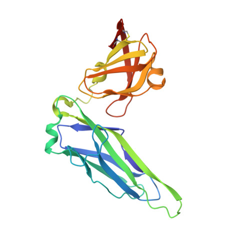
    2W07

  • PubMed Abstract: 

    P pili are important adhesive fibers involved in kidney infection by uropathogenic Escherichia coli. Pilus subunits are characterized by a large groove resulting from lack of a beta strand. Polymerization of pilus subunits occurs via the donor-strand exchange (DSE) mechanism initiated when the N terminus of an incoming subunit interacts with the P5 region/pocket of the previously assembled subunit groove. Here, we solve the structure of the PapD:PapF complex in order to understand why PapF undergoes slow DSE. The structure reveals that the PapF P5 pocket is partially obstructed. MD simulations show this region of PapF is flexible compared with its equivalent in PapH, a subunit that also has an obstructed P5 pocket and is unable to undergo DSE. Using electrospray-ionization mass spectrometry, we show that mutations in the P5 region result in increased DSE rates. Thus, partial obstruction of the P5 pocket serves as a modulating mechanism of DSE.


  • Organizational Affiliation
    • Institute of Structural and Molecular Biology, University College London, Birkbeck College, Malet Street, London WC1E 7HX, UK.

Macromolecule Content 

  • Total Structure Weight: 40.55 kDa 
  • Atom Count: 2,626 
  • Modeled Residue Count: 337 
  • Deposited Residue Count: 366 
  • Unique protein chains: 2

Macromolecules

Find similar proteins by:|  3D Structure
Entity ID: 1
MoleculeChains  Sequence LengthOrganismDetailsImage
CHAPERONE PROTEIN PAPD218Escherichia coli UTI89Mutation(s): 0 
UniProt
Find proteins for P15319 (Escherichia coli)
Explore P15319 
Go to UniProtKB:  P15319
Entity Groups
Sequence Clusters30% Identity50% Identity70% Identity90% Identity95% Identity100% Identity
UniProt GroupP15319
Sequence Annotations
Expand
Reference Sequence
Find similar proteins by:|  3D Structure
Entity ID: 2
MoleculeChains  Sequence LengthOrganismDetailsImage
MINOR PILIN SUBUNIT PAPF148Escherichia coli UTI89Mutation(s): 1 
UniProt
Find proteins for P42187 (Escherichia coli)
Explore P42187 
Go to UniProtKB:  P42187
Entity Groups
Sequence Clusters30% Identity50% Identity70% Identity90% Identity95% Identity100% Identity
UniProt GroupP42187
Sequence Annotations
Expand
Reference Sequence

Experimental Data & Validation

Experimental Data

  • Method: X-RAY DIFFRACTION
  • Resolution: 2.20 Å
  • R-Value Free:  0.277 (Depositor), 0.272 (DCC) 
  • R-Value Work:  0.235 (Depositor), 0.230 (DCC) 
  • R-Value Observed: 0.235 (Depositor) 
Space Group: P 41 21 2
Unit Cell:
Length ( Å )Angle ( ˚ )
a = 65.42α = 90
b = 65.42β = 90
c = 166.11γ = 90
Software Package:
Software NamePurpose
CNSrefinement
MOSFLMdata reduction
SCALAdata scaling
AMoREphasing

Structure Validation

View Full Validation Report


Entry History 

Deposition Data

Revision History  (Full details and data files)

  • Version 1.0: 2008-11-25
    Type: Initial release
  • Version 1.1: 2011-05-07
    Changes: Version format compliance
  • Version 1.2: 2011-07-13
    Changes: Version format compliance
  • Version 1.3: 2023-12-13
    Changes: Data collection, Database references, Other, Refinement description
  • Version 1.4: 2024-11-06
    Changes: Structure summary