On-Line Optical and X-Ray Spectroscopies with Crystallography: An Integrated Approach for Determining Metalloprotein Structures in Functionally Well Defined States.
Ellis, M.J., Buffey, S.G., Hough, M.A., Hasnain, S.S.(2008) J Synchrotron Radiat 15: 433
- PubMed: 18728313
- DOI: https://doi.org/10.1107/S0909049508014945
- Primary Citation of Related Structures:
2VW4, 2VW6, 2VW7 - PubMed Abstract:
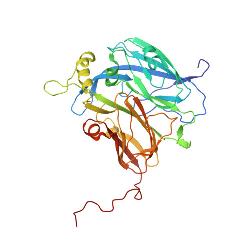
X-ray-induced redox changes can lead to incorrect assignments of the functional states of metals in metalloprotein crystals. The need for on-line monitoring of the status of metal ions (and other chromophores) during protein crystallography experiments is of growing importance with the use of intense synchrotron X-ray beams. Significant efforts are therefore being made worldwide to combine different spectroscopies in parallel with X-ray crystallographic data collection. Here the implementation and utilization of optical and X-ray absorption spectroscopies on the modern macromolecular crystallography (MX) beamline 10, at the SRS, Daresbury Laboratory, is described. This beamline is equipped with a dedicated monolithic energy-dispersive X-ray fluorescence detector, allowing X-ray absorption spectroscopy (XAS) measurements to be made in situ on the same crystal used to record the diffraction data. In addition, an optical microspectrophotometer has been incorporated on the beamline, thus facilitating combined MX, XAS and optical spectroscopic measurements. By uniting these techniques it is also possible to monitor the status of optically active and optically silent metal centres present in a crystal at the same time. This unique capability has been applied to observe the results of crystallographic data collection on crystals of nitrite reductase from Alcaligenes xylosoxidans, which contains both type-1 and type-2 Cu centres. It is found that the type-1 Cu centre photoreduces quickly, resulting in the loss of the 595 nm peak in the optical spectrum, while the type-2 Cu centre remains in the oxidized state over a much longer time period, for which independent confirmation is provided by XAS data as this centre has an optical spectrum which is barely detectable using microspectrophotometry. This example clearly demonstrates the importance of using two on-line methods, spectroscopy and XAS, for identifying well defined redox states of metalloproteins during crystallographic data collection.
- Molecular Biophysics Group, STFC Daresbury Laboratory, Warrington WA4 4AD, UK.
Organizational Affiliation:


