2VUE

Human serum albumin complexed with 4Z,15E-bilirubin-IX-alpha


Experimental Data Snapshot

  • Method: X-RAY DIFFRACTION
  • Resolution: 2.42 Å
  • R-Value Free: 0.273 
  • R-Value Work: 0.218 
  • R-Value Observed: 0.218 

wwPDB Validation   3D Report Full Report


Ligand Structure Quality Assessment 


This is version 1.4 of the entry. See complete history


Literature

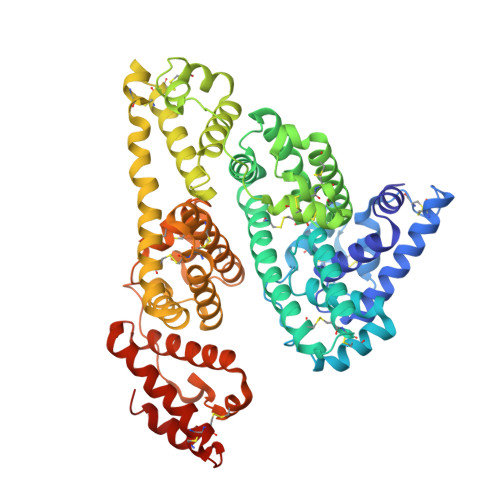
Crystallographic Analysis of Human Serum Albumin Complexed with 4Z,15E-Bilirubin-Ixalpha.

Zunszain, P.A.Ghuman, J.Mcdonagh, A.F.Curry, S.

(2008) J Mol Biol 381: 394

  • DOI: https://doi.org/10.1016/j.jmb.2008.06.016
  • Primary Citation of Related Structures:  
    2VUE, 2VUF

  • PubMed Abstract: 

    Bilirubin, an insoluble yellow-orange pigment derived from heme catabolism, accumulates to toxic levels in individuals with impaired or immature liver function. The resulting jaundice may be managed with phototherapy to isomerize the biosynthetic 4Z,15Z-bilirubin-IXalpha to more soluble and excretable isomers, such as 4Z,15E-bilirubin. Bilirubin and its configurational isomers are transported to the liver by human serum albumin (HSA) but their precise binding location(s) on the protein have yet to be determined. To investigate the molecular details of their interaction, we co-crystallised bilirubin with HSA. Strikingly, the crystal structure--determined to 2.42 A resolution--revealed the 4Z,15E-bilirubin-IXalpha isomer bound to an L-shaped pocket in sub-domain IB. We also determined the co-crystal structure of HSA complexed with fusidic acid, an antibiotic that competitively displaces bilirubin from the protein, and showed that it binds to the same pocket. These results provide the first crystal structure of a natural bilirubin pigment bound to serum albumin, challenge some of the present conceptions about HSA-bilirubin interactions, and provide a sound structural framework for finally resolving the long-standing question of where 4Z,15Z-bilirubin-IXalpha binds to the protein.


  • Organizational Affiliation

    Biophysics Section, Blackett Laboratory, Imperial College, South Kensington Campus, Exhibition Road, London SW7 2AZ, UK.


Macromolecules
Find similar proteins by:  (by identity cutoff)  |  3D Structure
Entity ID: 1
MoleculeChains Sequence LengthOrganismDetailsImage
SERUM ALBUMIN
A, B
585Homo sapiensMutation(s): 0 
UniProt & NIH Common Fund Data Resources
Find proteins for P02768 (Homo sapiens)
Explore P02768 
Go to UniProtKB:  P02768
PHAROS:  P02768
GTEx:  ENSG00000163631 
Entity Groups  
Sequence Clusters30% Identity50% Identity70% Identity90% Identity95% Identity100% Identity
UniProt GroupP02768
Sequence Annotations
Expand
  • Reference Sequence
Small Molecules
Experimental Data & Validation

Experimental Data

  • Method: X-RAY DIFFRACTION
  • Resolution: 2.42 Å
  • R-Value Free: 0.273 
  • R-Value Work: 0.218 
  • R-Value Observed: 0.218 
  • Space Group: P 1
Unit Cell:
Length ( Å )Angle ( ˚ )
a = 55.09α = 81.22
b = 55.54β = 90.7
c = 119.83γ = 65.42
Software Package:
Software NamePurpose
CNSrefinement
MOSFLMdata reduction
SCALAdata scaling
CNSphasing

Structure Validation

View Full Validation Report


Ligand Structure Quality Assessment 


Entry History 

Deposition Data

Revision History  (Full details and data files)

  • Version 1.0: 2008-06-17
    Type: Initial release
  • Version 1.1: 2011-05-08
    Changes: Version format compliance
  • Version 1.2: 2011-07-13
    Changes: Version format compliance
  • Version 1.3: 2019-07-24
    Changes: Data collection
  • Version 1.4: 2023-12-13
    Changes: Data collection, Database references, Other, Refinement description