Molecular Basis of Drug Resistance in Aurora Kinases.
Girdler, F., Sessa, F., Patercoli, S., Villa, F., Musacchio, A., Taylor, S.S.(2008) Chem Biol 15: 552
- PubMed: 18559266 Search on PubMed
- DOI: https://doi.org/10.1016/j.chembiol.2008.04.013
- Primary Citation Related Structures:
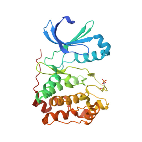
2VRX - PubMed Abstract:
Aurora kinases have emerged as potential targets in cancer therapy, and several drugs are currently undergoing preclinical and clinical validation. Whether clinical resistance to these drugs can arise is unclear. We exploited a hypermutagenic cancer cell line to select mutations conferring resistance to a well-studied Aurora inhibitor, ZM447439. All resistant clones contained dominant point mutations in Aurora B. Three mutations map to residues in the ATP-binding pocket that are distinct from the "gatekeeper" residue. The mutants retain wild-type catalytic activity and were resistant to all of the Aurora inhibitors tested. Our studies predict that drug-resistant Aurora B mutants are likely to arise during clinical treatment. Furthermore, because the plasticity of the ATP-binding pocket renders Aurora B insensitive to multiple inhibitors, our observations indicate that the drug-resistant Aurora B mutants should be exploited as novel drug targets.
- Faculty of Life Sciences, University of Manchester, Manchester, UK.
Organizational Affiliation: