Structural Insight Into Bioremediation of Triphenylmethane Dyes by Citrobacter Sp. Triphenylmethane Reductase.
Kim, M.H., Kim, Y., Park, H.J., Lee, J.S., Kwak, S.N., Jung, W.H., Lee, S.G., Kim, D., Lee, Y.C., Oh, T.K.(2008) J Biol Chem 283: 31981
- PubMed: 18782772 Search on PubMed
- DOI: https://doi.org/10.1074/jbc.M804092200
- Primary Citation Related Structures:
2JL1, 2VRB, 2VRC - PubMed Abstract:
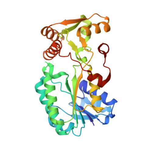
Triphenylmethane dyes are aromatic xenobiotic compounds that are widely considered to be one of the main culprits of environmental pollution. Triphenylmethane reductase (TMR) from Citrobacter sp. strain KCTC 18061P was initially isolated and biochemically characterized as an enzyme that catalyzes the reduction of triphenylmethane dyes. Information from the primary amino acid sequence suggests that TMR is a dinucleotide-binding motif-containing enzyme; however, no other functional clues can be derived from sequence analysis. We present the crystal structure of TMR in complex with NADP+ at 2.0-angstroms resolution. Despite limited sequence similarity, the enzyme shows remarkable structural similarity to short-chain dehydrogenase/reductase (SDR) family proteins. Functional assignments revealed that TMR has features of both classic and extended SDR family members and does not contain a conserved active site. Thus, it constitutes a novel class of SDR family proteins. On the basis of simulated molecular docking using the substrate malachite green and the TMR/NADP+ crystal structure, together with site-directed mutagenesis, we have elucidated a potential molecular mechanism for triphenylmethane dye reduction.
- Systems Microbiology Research Center, Korea Research Institute of Bioscience and Biotechnology, Daejeon 305-806. mhk8n@kribb.re.kr
Organizational Affiliation: