2VJJ

TAILSPIKE PROTEIN OF E.COLI BACTERIOPHAGE HK620 IN COMPLEX WITH HEXASACCHARIDE


Experimental Data Snapshot

  • Method: X-RAY DIFFRACTION
  • Resolution: 1.59 Å
  • R-Value Free: 0.165 
  • R-Value Work: 0.133 
  • R-Value Observed: 0.134 

wwPDB Validation   3D Report Full Report


This is version 2.1 of the entry. See complete history


Literature

Crystal Structure of Escherichia Coli Phage Hk620 Tailspike: Podoviral Tailspike Endoglycosidase Modules are Evolutionarily Related.

Barbirz, S.Mueller, J.J.Uetrecht, C.Clark, A.J.Heinemann, U.Seckler, R.

(2008) Mol Microbiol 69: 303

  • DOI: https://doi.org/10.1111/j.1365-2958.2008.06311.x
  • Primary Citation of Related Structures:  
    2VJI, 2VJJ

  • PubMed Abstract: 

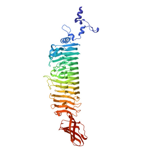
    Bacteriophage HK620 infects Escherichia coli H and is closely related to Shigella phage Sf6 and Salmonella phage P22. All three Podoviridae recognize and cleave their respective host cell receptor polysaccharide by homotrimeric tailspike proteins. The three proteins exhibit high sequence identity in the 110 residues of their N-terminal particle-binding domains, but no apparent sequence similarity in their major, receptor-binding parts. We have biochemically characterized the receptor-binding part of HK620 tailspike and determined its crystal structure to 1.38 A resolution. Its major domain is a right-handed parallel beta-helix, as in Sf6 and P22 tailspikes. HK620 tailspike has endo-N-acetylglucosaminidase activity and produces hexasaccharides of an O18A1-type O-antigen. As indicated by the structure of a hexasaccharide complex determined at 1.6 A resolution, the endoglycosidase-active sites are located intramolecularly, as in P22, and not between subunits, as in Sf6 tailspike. In contrast, the extreme C-terminal domain of HK620 tailspike forms a beta-sandwich, as in Sf6 and unlike P22 tailspike. Despite the different folds, structure-based sequence alignments of the C-termini reveal motifs conserved between the three proteins. We propose that the tailspike genes of P22, Sf6 and HK620 have a common precursor and are not mosaics of unrelated gene fragments.


  • Organizational Affiliation

    Physikalische Biochemie, Universität Potsdam, Karl-Liebknecht-Str. 24-25, 14476 Golm, Germany.


Macromolecules
Find similar proteins by:  (by identity cutoff)  |  3D Structure
Entity ID: 1
MoleculeChains Sequence LengthOrganismDetailsImage
TAILSPIKE PROTEIN600Enterobacteria phage HK620Mutation(s): 0 
UniProt
Find proteins for Q9AYY6 (Enterobacteria phage HK620)
Explore Q9AYY6 
Go to UniProtKB:  Q9AYY6
Entity Groups  
Sequence Clusters30% Identity50% Identity70% Identity90% Identity95% Identity100% Identity
UniProt GroupQ9AYY6
Sequence Annotations
Expand
  • Reference Sequence
Oligosaccharides

Help

Entity ID: 2
MoleculeChains Length2D Diagram Glycosylation3D Interactions
alpha-L-rhamnopyranose-(1-6)-alpha-D-glucopyranose-(1-4)-[2-acetamido-2-deoxy-beta-D-glucopyranose-(1-3)]beta-D-galactopyranose-(1-3)-[alpha-D-glucopyranose-(1-6)]2-acetamido-2-deoxy-beta-D-glucopyranose
B
6N/A
Glycosylation Resources
GlyTouCan:  G20402DC
GlyCosmos:  G20402DC
Small Molecules
Ligands 4 Unique
IDChains Name / Formula / InChI Key2D Diagram3D Interactions
GOL
Query on GOL

Download Ideal Coordinates CCD File 
F [auth A]
G [auth A]
H [auth A]
I [auth A]
J [auth A]
F [auth A],
G [auth A],
H [auth A],
I [auth A],
J [auth A],
K [auth A],
L [auth A]
GLYCEROL
C3 H8 O3
PEDCQBHIVMGVHV-UHFFFAOYSA-N
CA
Query on CA

Download Ideal Coordinates CCD File 
C [auth A]CALCIUM ION
Ca
BHPQYMZQTOCNFJ-UHFFFAOYSA-N
K
Query on K

Download Ideal Coordinates CCD File 
E [auth A]POTASSIUM ION
K
NPYPAHLBTDXSSS-UHFFFAOYSA-N
CL
Query on CL

Download Ideal Coordinates CCD File 
D [auth A]CHLORIDE ION
Cl
VEXZGXHMUGYJMC-UHFFFAOYSA-M
Experimental Data & Validation

Experimental Data

  • Method: X-RAY DIFFRACTION
  • Resolution: 1.59 Å
  • R-Value Free: 0.165 
  • R-Value Work: 0.133 
  • R-Value Observed: 0.134 
  • Space Group: P 3 2 1
Unit Cell:
Length ( Å )Angle ( ˚ )
a = 73.91α = 90
b = 73.91β = 90
c = 174.59γ = 120
Software Package:
Software NamePurpose
REFMACrefinement
XDSdata reduction
XSCALEdata scaling
REFMACphasing

Structure Validation

View Full Validation Report


Entry History 

Deposition Data

Revision History  (Full details and data files)

  • Version 1.0: 2008-07-01
    Type: Initial release
  • Version 1.1: 2015-01-21
    Changes: Atomic model, Database references, Derived calculations, Non-polymer description, Other, Source and taxonomy, Structure summary, Version format compliance
  • Version 1.2: 2019-01-30
    Changes: Data collection, Experimental preparation, Other
  • Version 1.3: 2019-02-06
    Changes: Data collection, Experimental preparation
  • Version 2.0: 2020-07-29
    Type: Remediation
    Reason: Carbohydrate remediation
    Changes: Advisory, Atomic model, Data collection, Derived calculations, Other, Structure summary
  • Version 2.1: 2023-12-13
    Changes: Data collection, Database references, Refinement description, Structure summary