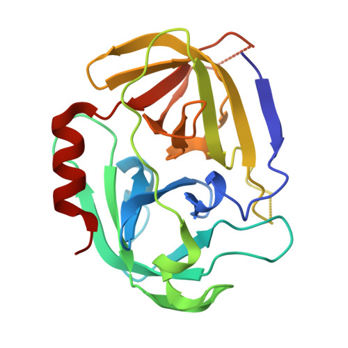
Enzymatic Activity of the Staphylococcus Aureus Splb Serine Protease is Induced by Substrates Containing the Sequence Trp-Glu-Leu-Gln.
Dubin, G., Stec-Niemczyk, J., Kisielewska, M., Pustelny, K., Popowicz, G.M., Bista, M., Kantyka, T., Boulware, K.T., Stennicke, H.R., Czarna, A., Phopaisarn, M., Daugherty, P.S., Thogersen, I.B., Enghild, J.J., Thornberry, N., Dubin, A., Potempa, J.(2008) J Mol Biology 379: 343
- PubMed: 18448121
- DOI: https://doi.org/10.1016/j.jmb.2008.03.059
- Primary Citation of Related Structures:
2VID - PubMed Abstract:
Proteases are of significant importance for the virulence of Staphylococcus aureus. Nevertheless, their subset, the serine protease-like proteins, remains poorly characterized. Here presented is an investigation of SplB protease catalytic activity revealing that the enzyme possesses exquisite specificity and only cleaves efficiently after the sequence Trp-Glu-Leu-Gln. To understand the molecular basis for such selectivity, we solved the three-dimensional structure of SplB to 1.8 A. Modeling of substrate binding to the protease demonstrated that selectivity relies in part on a canonical specificity pockets-based mechanism. Significantly, the conformation of residues that ordinarily form the oxyanion hole, an essential structural element of the catalytic machinery of serine proteases, is not canonical in the SplB structure. We postulate that within SplB, the oxyanion hole is only formed upon docking of a substrate containing the consensus sequence motif. It is suggested that this unusual activation mechanism is used in parallel with classical determinants to further limit enzyme specificity. Finally, to guide future development, we attempt to point at likely physiological substrates and thus the role of SplB in staphylococcal physiology.
- Department of Microbiology, Faculty of Biochemistry, Biophysics and Biotechnology, Jagiellonian University, Gronostajowa 7, 30-387 Krakow, Poland. gdubin@mol.uj.edu.pl
Organizational Affiliation:


