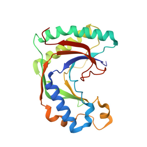
2VFK

AKAP18 delta central domain - AMP


Experimental Data Snapshot

  • Method: X-RAY DIFFRACTION
  • Resolution: 1.50 Å
  • R-Value Free: 0.209 
  • R-Value Work: 0.161 
  • R-Value Observed: 0.163 

Starting Model: other
View more details

wwPDB Validation   3D Report Full Report


Ligand Structure Quality Assessment 


This is version 1.3 of the entry. See complete history


Literature

Akap18 Contains a Phosphoesterase Domain that Binds AMP

Gold, M.G.Smith, F.D.Scott, J.D.Barford, D.

(2008) J Mol Biol 375: 1329

  • DOI: https://doi.org/10.1016/j.jmb.2007.11.037
  • Primary Citation of Related Structures:  
    2VFK, 2VFL, 2VFY

  • PubMed Abstract: 

    Protein kinase A anchoring proteins (AKAPs), defined by their capacity to target the cAMP-dependent protein kinase to distinct subcellular locations, function as molecular scaffolds mediating the assembly of multicomponent complexes to integrate and organise multiple signalling events. Despite their central importance in regulating cellular processes, little is known regarding their diverse structures and molecular mechanisms. Here, using bioinformatics and X-ray crystallography, we define a central domain of AKAP18 delta (AKAP18(CD)) as a member of the 2H phosphoesterase family. The domain features two conserved His-x-Thr motifs positioned at the base of a groove located between two lobes related by pseudo 2-fold symmetry. Nucleotide co-crystallisation screening revealed that this groove binds specifically to adenosine 5'-monophosphate (5'AMP) and cytosine 5'-monophosphate (5'CMP), with the affinity constant for AMP in the physiological concentration range. This is the first example of an AKAP capable of binding a small molecule. Our data generate two functional hypotheses for the AKAP18 central domain. It may act as a phosphoesterase, although we did not identify a substrate, or as an AMP sensor with the potential to couple intracellular AMP levels to PKA signalling events.


  • Organizational Affiliation

    Section of Structural Biology, Institute of Cancer Research, Chester Beatty Laboratories, 237 Fulham Road, London SW3 6JB, UK.


Macromolecules
Find similar proteins by:  (by identity cutoff)  |  3D Structure
Entity ID: 1
MoleculeChains Sequence LengthOrganismDetailsImage
AKAP18 DELTA205Homo sapiensMutation(s): 0 
Entity Groups  
Sequence Clusters30% Identity50% Identity70% Identity90% Identity95% Identity100% Identity
Sequence Annotations
Expand
  • Reference Sequence
Small Molecules
Ligands 1 Unique
IDChains Name / Formula / InChI Key2D Diagram3D Interactions
AMP
Query on AMP

Download Ideal Coordinates CCD File 
B [auth A]ADENOSINE MONOPHOSPHATE
C10 H14 N5 O7 P
UDMBCSSLTHHNCD-KQYNXXCUSA-N
Binding Affinity Annotations 
IDSourceBinding Affinity
AMP PDBBind:  2VFK Kd: 1.94e+5 (nM) from 1 assay(s)
Experimental Data & Validation

Experimental Data

  • Method: X-RAY DIFFRACTION
  • Resolution: 1.50 Å
  • R-Value Free: 0.209 
  • R-Value Work: 0.161 
  • R-Value Observed: 0.163 
  • Space Group: P 31
Unit Cell:
Length ( Å )Angle ( ˚ )
a = 42.233α = 90
b = 42.233β = 90
c = 97.723γ = 120
Software Package:
Software NamePurpose
REFMACrefinement
MOSFLMdata reduction
SCALAdata scaling
PHASERphasing

Structure Validation

View Full Validation Report


Ligand Structure Quality Assessment 


Entry History 

Deposition Data

Revision History  (Full details and data files)

  • Version 1.0: 2008-05-06
    Type: Initial release
  • Version 1.1: 2011-05-08
    Changes: Version format compliance
  • Version 1.2: 2011-07-13
    Changes: Version format compliance
  • Version 1.3: 2024-05-01
    Changes: Data collection, Database references, Other, Refinement description