2VD7 | pdb_00002vd7

Crystal Structure of JMJD2A complexed with inhibitor Pyridine-2,4- dicarboxylic acid


Experimental Data Snapshot

  • Method: X-RAY DIFFRACTION
  • Resolution: 2.25 Å
  • R-Value Free: 
    0.219 (Depositor), 0.213 (DCC) 
  • R-Value Work: 
    0.170 (Depositor), 0.168 (DCC) 
  • R-Value Observed: 
    0.172 (Depositor) 

wwPDB Validation 3D Report Full Report

Validation slider image for 2VD7

Ligand Structure Quality Assessment 


This is version 1.5 of the entry. See complete history

Literature

Inhibitor scaffolds for 2-oxoglutarate-dependent histone lysine demethylases.

Rose, N.R.Ng, S.S.Mecinovic, J.Lienard, B.M.Bello, S.H.Sun, Z.McDonough, M.A.Oppermann, U.Schofield, C.J.

(2008) J Med Chem 51: 7053-7056

  • DOI: https://doi.org/10.1021/jm800936s
  • Primary Citation Related Structures: 
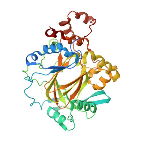
    2VD7

  • PubMed Abstract: 

    The dynamic methylation of histone lysyl residues plays an important role in biology by regulating transcription, maintaining genomic integrity, and by contributing to epigenetic effects. Here we describe a variety of inhibitor scaffolds that inhibit the human 2-oxoglutarate-dependent JMJD2 subfamily of histone demethylases. Combined with structural data, these chemical starting points will be useful to generate small-molecule probes to analyze the physiological roles of these enzymes in epigenetic signaling.


  • Organizational Affiliation
    • The Department of Chemistry and the Oxford Centre for Integrative Systems Biology, Chemistry Research Laboratory, University of Oxford, 12 Mansfield Road, Oxford, OX1 3TA, United Kingdom.

Macromolecule Content 

  • Total Structure Weight: 89.23 kDa 
  • Atom Count: 6,009 
  • Modeled Residue Count: 699 
  • Deposited Residue Count: 762 
  • Unique protein chains: 1

Macromolecules

Find similar proteins by:|  3D Structure
Entity ID: 1
MoleculeChains  Sequence LengthOrganismDetailsImage
JMJC DOMAIN-CONTAINING HISTONE DEMETHYLATION PROTEIN 3A
A, B
381Homo sapiensMutation(s): 0 
EC: 1.14.11 (PDB Primary Data), 1.14.11.66 (UniProt), 1.14.11.69 (UniProt)
UniProt & NIH Common Fund Data Resources
Find proteins for O75164 (Homo sapiens)
Explore O75164 
Go to UniProtKB:  O75164
PHAROS:  O75164
GTEx:  ENSG00000066135 
Entity Groups
Sequence Clusters30% Identity50% Identity70% Identity90% Identity95% Identity100% Identity
UniProt GroupO75164
Sequence Annotations
Expand
Reference Sequence

Small Molecules

Binding Affinity Annotations 
IDSourceBinding Affinity
PD2 BindingDB:  2VD7 IC50: 4200 (nM) from 1 assay(s)

Experimental Data & Validation

Experimental Data

  • Method: X-RAY DIFFRACTION
  • Resolution: 2.25 Å
  • R-Value Free:  0.219 (Depositor), 0.213 (DCC) 
  • R-Value Work:  0.170 (Depositor), 0.168 (DCC) 
  • R-Value Observed: 0.172 (Depositor) 
Space Group: P 21 21 2
Unit Cell:
Length ( Å )Angle ( ˚ )
a = 101.285α = 90
b = 149.121β = 90
c = 57.566γ = 90
Software Package:
Software NamePurpose
REFMACrefinement
MOSFLMdata reduction
SCALAdata scaling
REFMACphasing

Structure Validation

View Full Validation Report


Ligand Structure Quality Assessment 


Entry History 

Revision History  (Full details and data files)

  • Version 1.0: 2007-11-27
    Type: Initial release
  • Version 1.1: 2011-07-13
    Changes: Advisory, Version format compliance
  • Version 1.2: 2013-12-11
    Changes: Database references, Derived calculations
  • Version 1.3: 2018-01-24
    Changes: Source and taxonomy
  • Version 1.4: 2018-03-28
    Changes: Database references, Source and taxonomy
  • Version 1.5: 2024-05-08
    Changes: Data collection, Database references, Derived calculations, Other