2V8I

Structure of a Family 2 Pectate Lyase in a Native Form


Experimental Data Snapshot

  • Method: X-RAY DIFFRACTION
  • Resolution: 1.50 Å
  • R-Value Free: 0.243 
  • R-Value Work: 0.222 
  • R-Value Observed: 0.223 

wwPDB Validation   3D Report Full Report


This is version 1.2 of the entry. See complete history


Literature

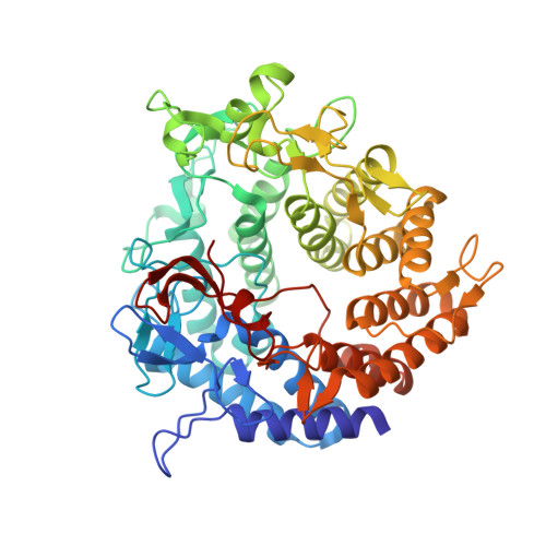
A Family 2 Pectate Lyase Displays a Rare Fold and Transition Metal-Assisted -Elimination.

Abbott, D.W.Boraston, A.B.

(2007) J Biol Chem 282: 35328

  • DOI: https://doi.org/10.1074/jbc.M705511200
  • Primary Citation of Related Structures:  
    2V8I, 2V8J, 2V8K

  • PubMed Abstract: 

    The family 2 pectate lyase from Yersinia enterocolitica (YePL2A), solved to 1.5A, reveals it to be the first prokaryotic protein reported to display the rare (alpha/alpha)(7) barrel fold. In addition to its apo form, we have also determined the structure of a metal-bound form of YePL2A (to 2.0A) and a trigalacturonic acid-bound substrate complex (to 2.1A) Although its fold is rare, the catalytic center of YePL2A can be superimposed with structurally unrelated families, underlining the conserved catalytic amino acid architecture of the beta-elimination mechanism. In addition to its overall structure, YePL2A also has two other unique features: 1) it utilizes a metal atom other than calcium for catalysis, and 2) its Brønstead base is in an alternate conformation and directly interacts with the uronate group of the substrate.


  • Organizational Affiliation

    Biochemistry & Microbiology, University of Victoria, Victoria, British Columbia V8W 3P6, Canada.


Macromolecules
Find similar proteins by:  (by identity cutoff)  |  3D Structure
Entity ID: 1
MoleculeChains Sequence LengthOrganismDetailsImage
PECTATE LYASE543Yersinia enterocoliticaMutation(s): 0 
EC: 4.2.2.2
UniProt
Find proteins for A1JSS8 (Yersinia enterocolitica serotype O:8 / biotype 1B (strain NCTC 13174 / 8081))
Explore A1JSS8 
Go to UniProtKB:  A1JSS8
Entity Groups  
Sequence Clusters30% Identity50% Identity70% Identity90% Identity95% Identity100% Identity
UniProt GroupA1JSS8
Sequence Annotations
Expand
  • Reference Sequence
Small Molecules
Ligands 1 Unique
IDChains Name / Formula / InChI Key2D Diagram3D Interactions
IOD
Query on IOD

Download Ideal Coordinates CCD File 
AA [auth A]
B [auth A]
BA [auth A]
C [auth A]
CA [auth A]
AA [auth A],
B [auth A],
BA [auth A],
C [auth A],
CA [auth A],
D [auth A],
DA [auth A],
E [auth A],
EA [auth A],
F [auth A],
FA [auth A],
G [auth A],
GA [auth A],
H [auth A],
HA [auth A],
I [auth A],
IA [auth A],
J [auth A],
JA [auth A],
K [auth A],
KA [auth A],
L [auth A],
LA [auth A],
M [auth A],
MA [auth A],
N [auth A],
NA [auth A],
O [auth A],
OA [auth A],
P [auth A],
PA [auth A],
Q [auth A],
QA [auth A],
R [auth A],
RA [auth A],
S [auth A],
SA [auth A],
T [auth A],
TA [auth A],
U [auth A],
UA [auth A],
V [auth A],
VA [auth A],
W [auth A],
WA [auth A],
X [auth A],
XA [auth A],
Y [auth A],
YA [auth A],
Z [auth A],
ZA [auth A]
IODIDE ION
I
XMBWDFGMSWQBCA-UHFFFAOYSA-M
Experimental Data & Validation

Experimental Data

  • Method: X-RAY DIFFRACTION
  • Resolution: 1.50 Å
  • R-Value Free: 0.243 
  • R-Value Work: 0.222 
  • R-Value Observed: 0.223 
  • Space Group: P 21 21 21
Unit Cell:
Length ( Å )Angle ( ˚ )
a = 58.486α = 90
b = 95.642β = 90
c = 126.608γ = 90
Software Package:
Software NamePurpose
REFMACrefinement

Structure Validation

View Full Validation Report


Entry History 

Deposition Data

Revision History  (Full details and data files)

  • Version 1.0: 2007-09-18
    Type: Initial release
  • Version 1.1: 2011-05-08
    Changes: Version format compliance
  • Version 1.2: 2011-07-13
    Changes: Version format compliance