Mechanism of enzymatic fluorination in Streptomyces cattleya.
Zhu, X., Robinson, D.A., McEwan, A.R., O'Hagan, D., Naismith, J.H.(2007) J Am Chem Soc 129: 14597-14604
- PubMed: 17985882 Search on PubMedSearch on PubMed Central
- DOI: https://doi.org/10.1021/ja0731569
- Primary Citation Related Structures:
2V7T, 2V7U, 2V7V, 2V7W, 2V7X - PubMed Abstract:
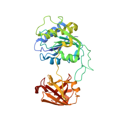
Recently a fluorination enzyme was identified and isolated from Streptomyces cattleya, as the first committed step on the metabolic pathway to the fluorinated metabolites, fluoroacetate and 4-fluorothreonine. This enzyme, 5'-fluoro-5'-deoxy adenosine synthetase (FDAS), has been shown to catalyze C-F bond formation by nucleophilic attack of fluoride ion to S-adenosyl-l-methionine (SAM) with the concomitant displacement of l-methionine to generate 5'-fluoro-5'-deoxy adenosine (5'-FDA). Although the structures of FDAS bound to both SAM and products have been solved, the molecular mechanism remained to be elucidated. We now report site-directed mutagenesis studies, structural analyses, and isothermal calorimetry (ITC) experiments. The data establish the key residues required for catalysis and the order of substrate binding. Fluoride ion is not readily distinguished from water by protein X-ray crystallography; however, using chloride ion (also a substrate) with a mutant of low activity has enabled the halide ion to be located in nonproductive co-complexes with SAH and SAM. The kinetic data suggest the positively charged sulfur of SAM is a key requirement in stabilizing the transition state. We propose a molecular mechanism for FDAS in which fluoride weakly associates with the enzyme exchanging two water molecules for protein ligation. The binding of SAM expels remaining water associated with fluoride ion and traps the ion in a pocket positioned to react with SAM, generating l-methionine and 5'-FDA. l-methionine then dissociates from the enzyme followed by 5'-FDA.
- Center for Biomolecular Sciences, University of St. Andrews, St. Andrews, KY16 9ST, United Kingdom.
Organizational Affiliation: