2V3Q

Serendipitous discovery and X-ray structure of a human phosphate binding apolipoprotein


Experimental Data Snapshot

  • Method: X-RAY DIFFRACTION
  • Resolution: 1.89 Å
  • R-Value Free: 0.186 
  • R-Value Work: 0.127 
  • R-Value Observed: 0.130 

wwPDB Validation   3D Report Full Report


This is version 1.1 of the entry. See complete history


Literature

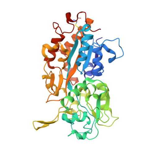
Tandem Use of X-Ray Crystallography and Mass Spectrometry to Obtain Ab Initio the Complete and Exact Amino Acids Sequence of Hpbp, a Human 38kDa Apolipoprotein

Diemer, H.Elias, M.Renault, F.Rochu, D.Contreras-Martel, C.Schaeffer, C.Van Dorsselaer, A.Chabriere, E.

(2008) Proteins 71: 1708

  • DOI: https://doi.org/10.1002/prot.21866
  • Primary Citation of Related Structures:  
    2V3Q

  • PubMed Abstract: 

    The Human Phosphate Binding Protein (HPBP) is a serendipitously discovered apolipoprotein from human plasma that binds phosphate. Amino acid sequence relates HPBP to an intriguing protein family that seems ubiquitous in eukaryotes. These proteins, named DING according to the sequence of their four conserved N-terminal residues, are systematically absent from eukaryotic genome databases. As a consequence, HPBP amino acids sequence had to be first assigned from the electronic density map. Then, an original approach combining X-ray crystallography and mass spectrometry provides the complete and a priori exact sequence of the 38-kDa HPBP. This first complete sequence of a eukaryotic DING protein will be helpful to study HPBP and the entire DING protein family.


  • Organizational Affiliation

    Laboratoire de Spectrométrie de Masse Bioorganique, Institut Pluridisciplinaire Hubert Curien, UMR 7178 (CNRS-ULP) ECPM, 25 rue Becquerel F67087-Strasbourg-Cedex 2, France.


Macromolecules
Find similar proteins by:  (by identity cutoff)  |  3D Structure
Entity ID: 1
MoleculeChains Sequence LengthOrganismDetailsImage
HUMAN PHOSPHATE BINDING PROTEIN376Homo sapiensMutation(s): 0 
UniProt
Find proteins for P85173 (Unknown prokaryotic organism)
Explore P85173 
Go to UniProtKB:  P85173
Entity Groups  
Sequence Clusters30% Identity50% Identity70% Identity90% Identity95% Identity100% Identity
UniProt GroupP85173
Sequence Annotations
Expand
  • Reference Sequence
Small Molecules
Ligands 3 Unique
IDChains Name / Formula / InChI Key2D Diagram3D Interactions
PO4
Query on PO4

Download Ideal Coordinates CCD File 
D [auth A]PHOSPHATE ION
O4 P
NBIIXXVUZAFLBC-UHFFFAOYSA-K
GOL
Query on GOL

Download Ideal Coordinates CCD File 
B [auth A],
C [auth A],
E [auth A],
P [auth A]
GLYCEROL
C3 H8 O3
PEDCQBHIVMGVHV-UHFFFAOYSA-N
EDO
Query on EDO

Download Ideal Coordinates CCD File 
F [auth A]
G [auth A]
H [auth A]
I [auth A]
J [auth A]
F [auth A],
G [auth A],
H [auth A],
I [auth A],
J [auth A],
K [auth A],
L [auth A],
M [auth A],
N [auth A],
O [auth A],
Q [auth A],
R [auth A]
1,2-ETHANEDIOL
C2 H6 O2
LYCAIKOWRPUZTN-UHFFFAOYSA-N
Experimental Data & Validation

Experimental Data

  • Method: X-RAY DIFFRACTION
  • Resolution: 1.89 Å
  • R-Value Free: 0.186 
  • R-Value Work: 0.127 
  • R-Value Observed: 0.130 
  • Space Group: C 2 2 21
Unit Cell:
Length ( Å )Angle ( ˚ )
a = 96.245α = 90
b = 86.843β = 90
c = 89.889γ = 90
Software Package:
Software NamePurpose
REFMACrefinement
XDSdata reduction
XDSdata scaling
SHARPphasing

Structure Validation

View Full Validation Report


Entry History 

Revision History  (Full details and data files)

  • Version 1.0: 2008-07-22
    Type: Initial release
  • Version 1.1: 2016-01-27
    Changes: Data collection, Derived calculations, Non-polymer description, Other, Version format compliance