2V1U | pdb_00002v1u

STRUCTURE OF THE AEROPYRUM PERNIX ORC1 PROTEIN IN COMPLEX WITH DNA


Experimental Data Snapshot

  • Method: X-RAY DIFFRACTION
  • Resolution: 3.10 Å
  • R-Value Free: 
    0.297 (Depositor), 0.267 (DCC) 
  • R-Value Work: 
    0.254 (Depositor), 0.255 (DCC) 
  • R-Value Observed: 
    0.257 (Depositor) 

wwPDB Validation 3D Report Full Report

Validation slider image for 2V1U

Ligand Structure Quality Assessment 


This is version 1.2 of the entry. See complete history

Literature

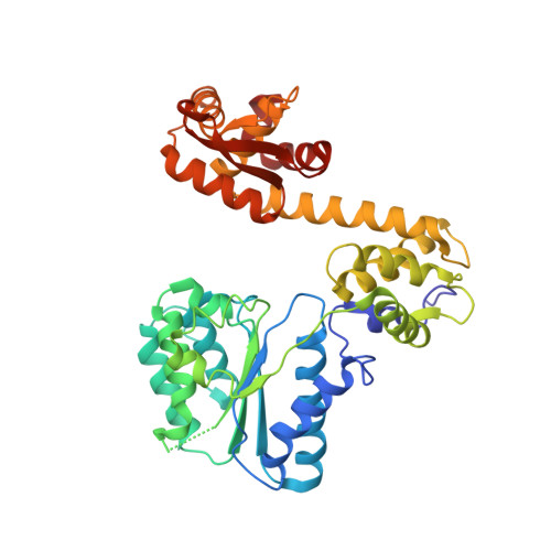
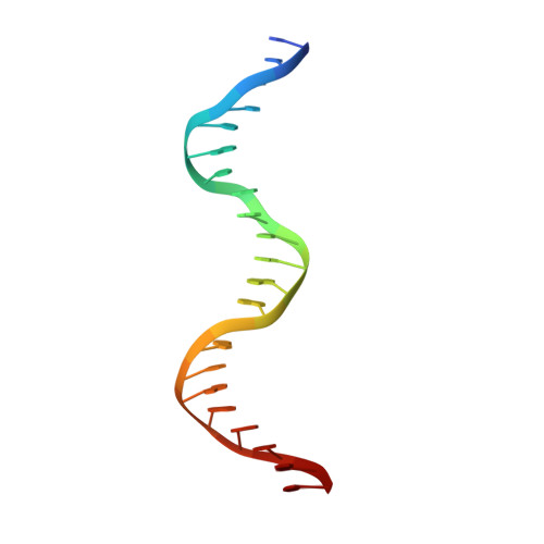
Structural Basis of DNA Replication Origin Recognition by an Orc Protein.

Gaudier, M.Schuwirth, B.S.Westcott, S.L.Wigley, D.B.

(2007) Science 317: 1213

  • DOI: https://doi.org/10.1126/science.1143664
  • Primary Citation Related Structures: 
    2V1U

  • PubMed Abstract: 

    DNA replication in archaea and in eukaryotes share many similarities. We report the structure of an archaeal origin recognition complex protein, ORC1, bound to an origin recognition box, a DNA sequence that is found in multiple copies at replication origins. DNA binding is mediated principally by a C-terminal winged helix domain that inserts deeply into the major and minor grooves, widening them both. However, additional DNA contacts are made with the N-terminal AAA+ domain, which inserts into the minor groove at a characteristic G-rich sequence, inducing a 35 degrees bend in the duplex and providing directionality to the binding site. Both contact regions also induce substantial unwinding of the DNA. The structure provides insight into the initial step in assembly of a replication origin and recruitment of minichromosome maintenance (MCM) helicase to that origin.


  • Organizational Affiliation
    • Cancer Research UK Clare Hall Laboratories, London Research Institute, Blanche Lane, South Mimms, Potters Bar, Herts EN6 3LD, UK.

Macromolecule Content 

  • Total Structure Weight: 57.46 kDa 
  • Atom Count: 3,951 
  • Modeled Residue Count: 426 
  • Deposited Residue Count: 431 
  • Unique protein chains: 1
  • Unique nucleic acid chains: 2

Macromolecules


Find similar proteins by:|  3D Structure
Entity ID: 1
MoleculeChains  Sequence LengthOrganismDetailsImage
CELL DIVISION CONTROL PROTEIN 6 HOMOLOG387Aeropyrum pernixMutation(s): 0 
UniProt
Find proteins for Q9YEV6 (Aeropyrum pernix (strain ATCC 700893 / DSM 11879 / JCM 9820 / NBRC 100138 / K1))
Explore Q9YEV6 
Go to UniProtKB:  Q9YEV6
Entity Groups
Sequence Clusters30% Identity50% Identity70% Identity90% Identity95% Identity100% Identity
UniProt GroupQ9YEV6
Sequence Annotations
Expand
Reference Sequence
Find similar nucleic acids by:  Sequence
Entity ID: 2
MoleculeChains LengthOrganismImage
5'-D(*TP*CP*TP*CP*CP*AP*CP*AP*GP*GP *AP*AP*AP*CP*GP*GP*AP*GP*GP*GP*GP*T)-3'22Aeropyrum pernix
Sequence Annotations
Expand
Reference Sequence
Find similar nucleic acids by:  Sequence
Entity ID: 3
MoleculeChains LengthOrganismImage
5'-D(*AP*CP*CP*CP*CP*TP*CP*CP*GP*TP *TP*TP*CP*CP*TP*GP*TP*GP*GP*AP*GP*A)-3'22Aeropyrum pernix
Sequence Annotations
Expand
Reference Sequence

Small Molecules

Experimental Data & Validation

Experimental Data

  • Method: X-RAY DIFFRACTION
  • Resolution: 3.10 Å
  • R-Value Free:  0.297 (Depositor), 0.267 (DCC) 
  • R-Value Work:  0.254 (Depositor), 0.255 (DCC) 
  • R-Value Observed: 0.257 (Depositor) 
Space Group: P 65 2 2
Unit Cell:
Length ( Å )Angle ( ˚ )
a = 75.7α = 90
b = 75.7β = 90
c = 395.11γ = 120
Software Package:
Software NamePurpose
MOSFLMdata reduction
SCALAdata scaling
PHASERphasing
autoSHARPphasing
REFMACrefinement

Structure Validation

View Full Validation Report


Ligand Structure Quality Assessment 


Entry History 

Deposition Data

Revision History  (Full details and data files)

  • Version 1.0: 2007-09-18
    Type: Initial release
  • Version 1.1: 2011-07-13
    Changes: Advisory, Version format compliance
  • Version 1.2: 2024-05-08
    Changes: Data collection, Database references, Derived calculations, Other