2UYF | pdb_00002uyf

Single mutant F111L DntR from Burkholderia sp. strain DNT in complex with thiocyanate


Experimental Data Snapshot

  • Method: X-RAY DIFFRACTION
  • Resolution: 2.20 Å
  • R-Value Free: 
    0.233 (Depositor), 0.233 (DCC) 
  • R-Value Work: 
    0.207 (Depositor), 0.206 (DCC) 
  • R-Value Observed: 
    0.208 (Depositor) 

Starting Model: experimental
View more details

wwPDB Validation 3D Report Full Report

Validation slider image for 2UYF

This is version 1.3 of the entry. See complete history

Literature

In Vivo and in Vitro Investigation of Transcriptional Regulation by Dntr.

Lonneborg, R.Smirova, I.Dian, C.Leonard, G.A.Brzezinski, P.

(2007) J Mol Biology 372: 571

  • DOI: https://doi.org/10.1016/j.jmb.2007.06.076
  • Primary Citation Related Structures: 
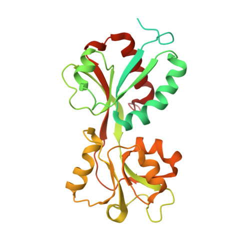
    2UYE, 2UYF

  • PubMed Abstract: 

    DntR is a bacterial transcription factor that has been isolated from Burkholderia species that are able to degrade the nitro-aromatic compound 2,4-dinitrotoluene. We recently solved the X-ray crystal structure of DntR, which suggested a putative location of an inducer-binding cavity (IBC). In this study, we constructed mutants of DntR in which residues lining the proposed IBC were modified in order to identify the structural elements involved in inducer binding, to modulate the inducer binding specificity, and to investigate the mechanism of transcriptional regulation by DntR. The transcriptional activation of the reporter gene gfp induced by the wild-type and mutant DntRs was monitored by analysing whole-cell fluorescence using flow-cytometry after addition of a number of potential inducer compounds. Three of the mutant proteins (F111L; F111V/H169V and Y110S/F111V) were purified and the binding constants for several of the potential inducers to these mutants were estimated. Furthermore, crystal structures of the F111L and Y110S/F111V mutant proteins were solved and used to explain changes in the inducer binding specificity at an atomic level. A comparison of the inducing capability in the whole-cell system and binding constants for a number of potential inducers suggests a mechanism where binding of an inducer molecule is not the sole requirement for transcriptional activation. In addition, specific interactions between DntR and the inducer molecule resulting in a conformational change of the protein are needed.


  • Organizational Affiliation
    • Department of Biochemistry and Biophysics, Arrhenius Laboratories for Natural Sciences, Stockholm University, SE-106 91 Stockholm, Sweden.

Macromolecule Content 

  • Total Structure Weight: 70.49 kDa 
  • Atom Count: 3,717 
  • Modeled Residue Count: 441 
  • Deposited Residue Count: 614 
  • Unique protein chains: 1

Macromolecules

Find similar proteins by:|  3D Structure
Entity ID: 1
MoleculeChains  Sequence LengthOrganismDetailsImage
REGULATORY PROTEIN
A, B
307Burkholderia cepaciaMutation(s): 1 
UniProt
Find proteins for Q8VUD7 (Burkholderia cepacia)
Explore Q8VUD7 
Go to UniProtKB:  Q8VUD7
Entity Groups
Sequence Clusters30% Identity50% Identity70% Identity90% Identity95% Identity100% Identity
UniProt GroupQ8VUD7
Sequence Annotations
Expand
Reference Sequence

Small Molecules

Ligands 3 Unique
IDChains Name / Formula / InChI Key2D Diagram3D Interactions
GOL

Query on GOL


Download:Ideal Coordinates CCD File
K [auth A],
L [auth A],
Q [auth B],
R [auth B]
GLYCEROL
C3 H8 O3
PEDCQBHIVMGVHV-UHFFFAOYSA-N
ACT

Query on ACT


Download:Ideal Coordinates CCD File
S [auth B]ACETATE ION
C2 H3 O2
QTBSBXVTEAMEQO-UHFFFAOYSA-M
SCN

Query on SCN


Download:Ideal Coordinates CCD File
C [auth A]
D [auth A]
E [auth A]
F [auth A]
G [auth A]
C [auth A],
D [auth A],
E [auth A],
F [auth A],
G [auth A],
H [auth A],
I [auth A],
J [auth A],
M [auth B],
N [auth B],
O [auth B],
P [auth B]
THIOCYANATE ION
C N S
ZMZDMBWJUHKJPS-UHFFFAOYSA-M

Experimental Data & Validation

Experimental Data

  • Method: X-RAY DIFFRACTION
  • Resolution: 2.20 Å
  • R-Value Free:  0.233 (Depositor), 0.233 (DCC) 
  • R-Value Work:  0.207 (Depositor), 0.206 (DCC) 
  • R-Value Observed: 0.208 (Depositor) 
Space Group: P 65 2 2
Unit Cell:
Length ( Å )Angle ( ˚ )
a = 106.604α = 90
b = 106.604β = 90
c = 296.448γ = 120
Software Package:
Software NamePurpose
REFMACrefinement
XDSdata reduction
XDSdata scaling
REFMACphasing

Structure Validation

View Full Validation Report


Entry History 

Deposition Data

Revision History  (Full details and data files)

  • Version 1.0: 2007-08-21
    Type: Initial release
  • Version 1.1: 2011-05-08
    Changes: Version format compliance
  • Version 1.2: 2011-07-13
    Changes: Version format compliance
  • Version 1.3: 2023-12-13
    Changes: Data collection, Database references, Other, Refinement description