2TCL | pdb_00002tcl

STRUCTURE OF THE CATALYTIC DOMAIN OF HUMAN FIBROBLAST COLLAGENASE COMPLEXED WITH AN INHIBITOR


Experimental Data Snapshot

  • Method: X-RAY DIFFRACTION
  • Resolution: 2.20 Å
  • R-Value Work: 
    0.162 (Depositor) 
  • R-Value Observed: 
    0.162 (Depositor) 

wwPDB Validation 3D Report Full Report

Validation slider image for 2TCL

This is version 1.3 of the entry. See complete history

Literature

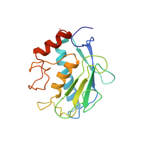
Structure of the catalytic domain of human fibroblast collagenase complexed with an inhibitor.

Borkakoti, N.Winkler, F.K.Williams, D.H.D'Arcy, A.Broadhurst, M.J.Brown, P.A.Johnson, W.H.Murray, E.J.

(1994) Nat Struct Biol 1: 106-110

  • DOI: https://doi.org/10.1038/nsb0294-106
  • Primary Citation Related Structures: 
    2TCL

  • PubMed Abstract: 

    In rheumatoid and osteoarthritis, degradation of articular cartilage is mediated by the matrix metalloproteinases collagenase, stromelysin and gelatinase. The key event in this process is the cleavage of triple helical collagen by collagenase. We have determined the crystal structure of the catalytic domain of human recombinant fibroblast collagenase complexed with a synthetic inhibitor at 2.2 A resolution. The protein fold is similar to the amino termini of the zinc endopeptidases astacin thermolysin and elastase despite a lack of primary sequence homology. The conformation of the bound inhibitor provides a molecular basis for the design of inhibitors of collagenase and other matrix metalloproteinases. Such inhibitors should be useful in the treatment of a variety of diseases including arthritis and cancer.


  • Organizational Affiliation
    • Roche Products Ltd, Garden City, Hertfordshire, UK.

Macromolecule Content 

  • Total Structure Weight: 19.63 kDa 
  • Atom Count: 1,387 
  • Modeled Residue Count: 163 
  • Deposited Residue Count: 169 
  • Unique protein chains: 1

Macromolecules

Find similar proteins by:|  3D Structure
Entity ID: 1
MoleculeChains  Sequence LengthOrganismDetailsImage
FIBROBLAST COLLAGENASE169Homo sapiensMutation(s): 0 
EC: 3.4.24.7
UniProt & NIH Common Fund Data Resources
Find proteins for P03956 (Homo sapiens)
Explore P03956 
Go to UniProtKB:  P03956
PHAROS:  P03956
GTEx:  ENSG00000196611 
Entity Groups
Sequence Clusters30% Identity50% Identity70% Identity90% Identity95% Identity100% Identity
UniProt GroupP03956
Sequence Annotations
Expand
Reference Sequence

Experimental Data & Validation

Experimental Data

  • Method: X-RAY DIFFRACTION
  • Resolution: 2.20 Å
  • R-Value Work:  0.162 (Depositor) 
  • R-Value Observed: 0.162 (Depositor) 
Space Group: I 4
Unit Cell:
Length ( Å )Angle ( ˚ )
a = 86.96α = 90
b = 86.96β = 90
c = 40.96γ = 90
Software Package:
Software NamePurpose
X-PLORmodel building
X-PLORrefinement
X-PLORphasing

Structure Validation

View Full Validation Report


Entry History 

Deposition Data

Revision History  (Full details and data files)

  • Version 1.0: 1996-03-08
    Type: Initial release
  • Version 1.1: 2008-03-25
    Changes: Version format compliance
  • Version 1.2: 2011-07-13
    Changes: Version format compliance
  • Version 1.3: 2024-02-21
    Changes: Data collection, Database references, Derived calculations, Other