2RTM

STREPTAVIDIN-2-IMINOBIOTIN-SULFATE COMPLEX, PH 3.50, SPACE GROUP I4122


Experimental Data Snapshot

  • Method: X-RAY DIFFRACTION
  • Resolution: 1.30 Å
  • R-Value Free: 0.238 
  • R-Value Work: 0.199 
  • R-Value Observed: 0.199 

wwPDB Validation   3D Report Full Report


Ligand Structure Quality Assessment 


This is version 1.3 of the entry. See complete history


Literature

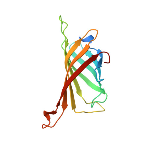
Binding of biotin to streptavidin stabilizes intersubunit salt bridges between Asp61 and His87 at low pH.

Katz, B.A.

(1997) J Mol Biol 274: 776-800

  • DOI: https://doi.org/10.1006/jmbi.1997.1444
  • Primary Citation of Related Structures:  
    2IZA, 2IZB, 2IZC, 2IZD, 2IZE, 2IZF, 2IZG, 2IZH, 2IZI, 2IZJ, 2IZK, 2IZL, 2RTA, 2RTB, 2RTC, 2RTD, 2RTE, 2RTF, 2RTG, 2RTH, 2RTI, 2RTJ, 2RTK, 2RTL, 2RTM, 2RTN, 2RTO, 2RTP, 2RTQ, 2RTR

  • PubMed Abstract: 
  • The remarkable stability of the streptavidin tetramer towards subunit dissociation becomes even greater upon binding of biotin. At two equivalent extensive monomer-monomer interfaces, monomers tightly associate into dimers that in turn associate into the tetramer at a less extensive dimer-dimer interface ...

    The remarkable stability of the streptavidin tetramer towards subunit dissociation becomes even greater upon binding of biotin. At two equivalent extensive monomer-monomer interfaces, monomers tightly associate into dimers that in turn associate into the tetramer at a less extensive dimer-dimer interface. To probe the structural basis for the enhancement of the stability of streptavidin by biotin, the crystal structures of apostreptavidin and its complexes with biotin and other small molecule and cyclic peptide ligands were determined and compared at resolutions as high as 1.36 A over a range of pH values from as low as 1.39. At low pH dramatic changes occur in the conformation and intersubunit hydrogen bonds involving the loop comprising Asp61 to Ser69. The hydrogen-bonded salt bridge between Asp61 Odelta2 and His87 Ndelta1, observed at higher pH, is replaced with a strong hydrogen bond between Asp61 Odelta1 and Asn85 Odelta1. Through crystallography at multiple pH values, the pH where this conformational change occurs, and thus the pKa of Asp61, was determined in crystals of space group I222 and/or I4122 of apostreptavidin and complexes. A range in pKa values for Asp61 was observed in these structures, the lowest being 1.78+/-0.19 for I222 streptavidin-biotin in 2.9 M (NH4)2SO4. At low pH the decrease in pKa of Asp61 and preservation of the intersubunit Asp61 Odelta2-Ndelta1 His87 hydrogen-bonded salt bridge in streptavidin-biotin versus apostreptavidin or streptavidin-peptide complexes is associated with an ordering of the flexible flap comprising residues Ala46 to Glu51, that in turn orders the Arg84 side-chain of a neighboring loop through resulting hydrogen bonds. Ordering of Arg84 in close proximity to the strong intersubunit interface appears to stabilize the conformation associated with the Asp61 Odelta2-Ndelta1 His87 hydrogen-bonded salt bridge. Thus, in addition to the established role of biotin in tetramer stabilization by direct mediation of intersubunit interactions at the weak interface through contact with Trp120, biotin may enhance tetramer stability at the strong interface more indirectly by ordering loop residues.


    Related Citations: 
    • In Crystals of Complexes of Streptavidin with Peptide Ligands Containing the Hpq Sequence the Pka of the Peptide Histidine is Less Than 3.0
      Katz, B.A., Cass, R.T.
      (1997) J Biol Chem 272: 13220
    • Structure-Based Design Tools: Structural and Thermodynamic Comparison with Biotin of a Small Molecule that Binds Streptavidin with Micromolar Affinity
      Katz, B.A., Liu, B., Cass, R.T.
      (1996) J Am Chem Soc 118: 7914
    • Preparation of a Protein-Dimerizing Ligand by Topochemistry and Structure-Based Design
      Katz, B.A.
      (1996) J Am Chem Soc 118: 2535
    • Topochemical Catalysis Achieved by Structure-Based Ligand Design
      Katz, B.A., Cass, R.T., Liu, B., Arze, R., Collins, N.
      (1995) J Biol Chem 270: 31210
    • Topochemistry for Preparing Ligands that Dimerize Receptors
      Katz, B.A., Stroud, R.M., Collins, N., Liu, B., Arze, R.
      (1995) Chem Biol 2: 591
    • Binding to Protein Targets of Peptidic Leads Discovered by Phage Display: Crystal Structures of Streptavidin-Bound Linear and Cyclic Peptide Ligands Containing the Hpq Sequence
      Katz, B.A.
      (1995) Biochemistry 34: 15421
    • Structure-Based Design of High Affinity Streptavidin Binding Cyclic Peptide Ligands Containing Thioether Cross-Links
      Katz, B.A., Johnson, C.R., Cass, R.T.
      (1995) J Am Chem Soc 117: 8541

    Organizational Affiliation

    Arris Pharmaceutical Corporation, 385 Oyster Point Boulevard, South San Francisco, CA 94080, USA.


Macromolecules
Find similar proteins by:  (by identity cutoff)  |  3D Structure
Entity ID: 1
MoleculeChainsSequence LengthOrganismDetailsImage
STREPTAVIDIN135Streptomyces avidiniiMutation(s): 0 
UniProt
Find proteins for P22629 (Streptomyces avidinii)
Explore P22629 
Go to UniProtKB:  P22629
Entity Groups  
Sequence Clusters30% Identity50% Identity70% Identity90% Identity95% Identity100% Identity
UniProt GroupP22629
Protein Feature View
Expand
  • Reference Sequence
Small Molecules
Ligands 2 Unique
IDChainsName / Formula / InChI Key2D Diagram3D Interactions
IMI
Query on IMI

Download Ideal Coordinates CCD File 
C [auth A]2-IMINOBIOTIN
C10 H17 N3 O2 S
WWVANQJRLPIHNS-ZKWXMUAHSA-N
 Ligand Interaction
SO4
Query on SO4

Download Ideal Coordinates CCD File 
B [auth A]SULFATE ION
O4 S
QAOWNCQODCNURD-UHFFFAOYSA-L
 Ligand Interaction
Experimental Data & Validation

Experimental Data

  • Method: X-RAY DIFFRACTION
  • Resolution: 1.30 Å
  • R-Value Free: 0.238 
  • R-Value Work: 0.199 
  • R-Value Observed: 0.199 
  • Space Group: I 41 2 2
Unit Cell:
Length ( Å )Angle ( ˚ )
a = 58.17α = 90
b = 58.17β = 90
c = 174.46γ = 90
Software Package:
Software NamePurpose
X-PLORmodel building
X-PLORrefinement
BIOTEXdata reduction
X-PLORphasing

Structure Validation

View Full Validation Report


Ligand Structure Quality Assessment 


Entry History 

Deposition Data

  • Released Date: 1998-10-14 
  • Deposition Author(s): Katz, B.A.

Revision History  (Full details and data files)

  • Version 1.0: 1998-10-14
    Type: Initial release
  • Version 1.1: 2008-03-25
    Changes: Version format compliance
  • Version 1.2: 2011-07-13
    Changes: Atomic model, Derived calculations, Version format compliance
  • Version 1.3: 2011-11-16
    Changes: Atomic model