2RIF | pdb_00002rif

CBS domain protein PAE2072 from Pyrobaculum aerophilum complexed with AMP


Experimental Data Snapshot

  • Method: X-RAY DIFFRACTION
  • Resolution: 2.35 Å
  • R-Value Free: 
    0.294 (Depositor), 0.290 (DCC) 
  • R-Value Work: 
    0.239 (Depositor), 0.237 (DCC) 
  • R-Value Observed: 
    0.242 (Depositor) 

wwPDB Validation 3D Report Full Report

Validation slider image for 2RIF

Ligand Structure Quality Assessment 


This is version 1.3 of the entry. See complete history

Literature

Structures and Functional Implications of an AMP-Binding Cystathionine beta-Synthase Domain Protein from a Hyperthermophilic Archaeon.

King, N.P.Lee, T.M.Sawaya, M.R.Cascio, D.Yeates, T.O.

(2008) J Mol Biology 380: 181-192

  • DOI: https://doi.org/10.1016/j.jmb.2008.04.073
  • Primary Citation Related Structures: 
    2RIF, 2RIH

  • PubMed Abstract: 

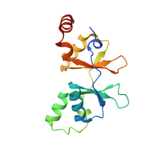
    Cystathionine beta-synthase domains are found in a myriad of proteins from organisms across the tree of life and have been hypothesized to function as regulatory modules that sense the energy charge of cells. Here we characterize the structure and stability of PAE2072, a dimeric tandem cystathionine beta-synthase domain protein from the hyperthermophilic crenarchaeon Pyrobaculum aerophilum. Crystal structures of the protein in unliganded and AMP-bound forms, determined at resolutions of 2.10 and 2.35 A, respectively, reveal remarkable conservation of key functional features seen in the gamma subunit of the eukaryotic AMP-activated protein kinase. The structures also confirm the presence of a suspected intermolecular disulfide bond between the two subunits that is shown to stabilize the protein. Our AMP-bound structure represents a first step in investigating the function of a large class of uncharacterized prokaryotic proteins. In addition, this work extends previous studies that have suggested that, in certain thermophilic microbes, disulfide bonds play a key role in stabilizing intracellular proteins and protein-protein complexes.


  • Organizational Affiliation
    • Department of Chemistry and Biochemistry, University of California, Los Angeles, Los Angeles, CA 90095, USA.

Macromolecule Content 

  • Total Structure Weight: 65.48 kDa 
  • Atom Count: 4,245 
  • Modeled Residue Count: 518 
  • Deposited Residue Count: 564 
  • Unique protein chains: 1

Macromolecules

Find similar proteins by:|  3D Structure
Entity ID: 1
MoleculeChains  Sequence LengthOrganismDetailsImage
Conserved protein with 2 CBS domains
A, B, C, D
141Pyrobaculum aerophilumMutation(s): 0 
Gene Names: PAE2072
UniProt
Find proteins for Q8ZVX8 (Pyrobaculum aerophilum (strain ATCC 51768 / DSM 7523 / JCM 9630 / CIP 104966 / NBRC 100827 / IM2))
Explore Q8ZVX8 
Go to UniProtKB:  Q8ZVX8
Entity Groups
Sequence Clusters30% Identity50% Identity70% Identity90% Identity95% Identity100% Identity
UniProt GroupQ8ZVX8
Sequence Annotations
Expand
Reference Sequence

Small Molecules

Ligands 2 Unique
IDChains Name / Formula / InChI Key2D Diagram3D Interactions
AMP

Query on AMP


Download:Ideal Coordinates CCD File
G [auth A]
H [auth A]
M [auth B]
N [auth B]
R [auth C]
G [auth A],
H [auth A],
M [auth B],
N [auth B],
R [auth C],
S [auth C],
V [auth D],
W [auth D]
ADENOSINE MONOPHOSPHATE
C10 H14 N5 O7 P
UDMBCSSLTHHNCD-KQYNXXCUSA-N
CS

Query on CS


Download:Ideal Coordinates CCD File
E [auth A]
F [auth A]
I [auth B]
J [auth B]
K [auth B]
E [auth A],
F [auth A],
I [auth B],
J [auth B],
K [auth B],
L [auth B],
O [auth C],
P [auth C],
Q [auth C],
T [auth D],
U [auth D]
CESIUM ION
Cs
NCMHKCKGHRPLCM-UHFFFAOYSA-N

Experimental Data & Validation

Experimental Data

  • Method: X-RAY DIFFRACTION
  • Resolution: 2.35 Å
  • R-Value Free:  0.294 (Depositor), 0.290 (DCC) 
  • R-Value Work:  0.239 (Depositor), 0.237 (DCC) 
  • R-Value Observed: 0.242 (Depositor) 
Space Group: P 41 21 2
Unit Cell:
Length ( Å )Angle ( ˚ )
a = 55.726α = 90
b = 55.726β = 90
c = 336.351γ = 90
Software Package:
Software NamePurpose
DENZOdata reduction
SCALEPACKdata scaling
PHASERphasing
REFMACrefinement
PDB_EXTRACTdata extraction

Structure Validation

View Full Validation Report


Ligand Structure Quality Assessment 


Entry History 

Deposition Data

Revision History  (Full details and data files)

  • Version 1.0: 2008-06-17
    Type: Initial release
  • Version 1.1: 2011-07-13
    Changes: Advisory, Version format compliance
  • Version 1.2: 2017-10-25
    Changes: Refinement description
  • Version 1.3: 2024-11-20
    Changes: Data collection, Database references, Derived calculations, Refinement description, Structure summary