2RHK

Crystal structure of influenza A NS1A protein in complex with F2F3 fragment of human cellular factor CPSF30, Northeast Structural Genomics Targets OR8C and HR6309A


Experimental Data Snapshot

  • Method: X-RAY DIFFRACTION
  • Resolution: 1.95 Å
  • R-Value Free: 0.234 
  • R-Value Work: 0.210 
  • R-Value Observed: 0.210 

wwPDB Validation   3D Report Full Report


This is version 1.2 of the entry. See complete history


Literature

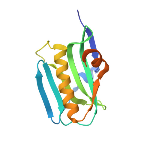
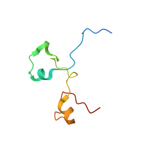
Structural basis for suppression of a host antiviral response by influenza A virus.

Das, K.Ma, L.C.Xiao, R.Radvansky, B.Aramini, J.Zhao, L.Marklund, J.Kuo, R.L.Twu, K.Y.Arnold, E.Krug, R.M.Montelione, G.T.

(2008) Proc Natl Acad Sci U S A 105: 13093-13098

  • DOI: https://doi.org/10.1073/pnas.0805213105
  • Primary Citation of Related Structures:  
    2RHK

  • PubMed Abstract: 
  • Influenza A viruses are responsible for seasonal epidemics and high mortality pandemics. A major function of the viral NS1A protein, a virulence factor, is the inhibition of the production of IFN-beta mRNA and other antiviral mRNAs. The NS1A protein of the human influenza A/Udorn/72 (Ud) virus inhibits the production of these antiviral mRNAs by binding the cellular 30-kDa subunit of the cleavage and polyadenylation specificity factor (CPSF30), which is required for the 3' end processing of all cellular pre-mRNAs ...

    Influenza A viruses are responsible for seasonal epidemics and high mortality pandemics. A major function of the viral NS1A protein, a virulence factor, is the inhibition of the production of IFN-beta mRNA and other antiviral mRNAs. The NS1A protein of the human influenza A/Udorn/72 (Ud) virus inhibits the production of these antiviral mRNAs by binding the cellular 30-kDa subunit of the cleavage and polyadenylation specificity factor (CPSF30), which is required for the 3' end processing of all cellular pre-mRNAs. Here we report the 1.95-A resolution X-ray crystal structure of the complex formed between the second and third zinc finger domain (F2F3) of CPSF30 and the C-terminal domain of the Ud NS1A protein. The complex is a tetramer, in which each of two F2F3 molecules wraps around two NS1A effector domains that interact with each other head-to-head. This structure identifies a CPSF30 binding pocket on NS1A comprised of amino acid residues that are highly conserved among human influenza A viruses. Single amino acid changes within this binding pocket eliminate CPSF30 binding, and a recombinant Ud virus expressing an NS1A protein with such a substitution is attenuated and does not inhibit IFN-beta pre-mRNA processing. This binding pocket is a potential target for antiviral drug development. The crystal structure also reveals that two amino acids outside of this pocket, F103 and M106, which are highly conserved (>99%) among influenza A viruses isolated from humans, participate in key hydrophobic interactions with F2F3 that stabilize the complex.


    Organizational Affiliation

    Center for Advanced Biotechnology and Medicine, Rutgers University, Piscataway, NJ 08854, USA.


Macromolecules
Find similar proteins by:  (by identity cutoff)  |  3D Structure
Entity ID: 1
MoleculeChainsSequence LengthOrganismDetailsImage
Non-structural protein 1
A, B
140Influenza A virusMutation(s): 0 
UniProt
Find proteins for P03495 (Influenza A virus (strain A/Udorn/307/1972 H3N2))
Explore P03495 
Go to UniProtKB:  P03495
Entity Groups  
Sequence Clusters30% Identity50% Identity70% Identity90% Identity95% Identity100% Identity
UniProt GroupP03495
Protein Feature View
Expand
  • Reference Sequence
Find similar proteins by:  (by identity cutoff)  |  3D Structure
Entity ID: 2
MoleculeChainsSequence LengthOrganismDetailsImage
Cleavage and polyadenylation specificity factor subunit 4
C, D
72Homo sapiensMutation(s): 1 
Gene Names: CPSF4CPSF30NARNEB1
UniProt & NIH Common Fund Data Resources
Find proteins for O95639 (Homo sapiens)
Explore O95639 
Go to UniProtKB:  O95639
PHAROS:  O95639
Entity Groups  
Sequence Clusters30% Identity50% Identity70% Identity90% Identity95% Identity100% Identity
UniProt GroupO95639
Protein Feature View
Expand
  • Reference Sequence
Experimental Data & Validation

Experimental Data

  • Method: X-RAY DIFFRACTION
  • Resolution: 1.95 Å
  • R-Value Free: 0.234 
  • R-Value Work: 0.210 
  • R-Value Observed: 0.210 
  • Space Group: P 41
Unit Cell:
Length ( Å )Angle ( ˚ )
a = 50.96α = 90
b = 50.96β = 90
c = 205.39γ = 90
Software Package:
Software NamePurpose
CNSrefinement
HKL-2000data reduction
SCALEPACKdata scaling
SOLVEphasing

Structure Validation

View Full Validation Report


Entry History 

Revision History  (Full details and data files)

  • Version 1.0: 2008-07-01
    Type: Initial release
  • Version 1.1: 2011-07-13
    Changes: Version format compliance
  • Version 1.2: 2021-10-20
    Changes: Database references, Derived calculations