2RG8

Crystal Structure of Programmed for Cell Death 4 Middle MA3 domain


Experimental Data Snapshot

  • Method: X-RAY DIFFRACTION
  • Resolution: 1.80 Å
  • R-Value Free: 0.251 
  • R-Value Work: 0.202 
  • R-Value Observed: 0.202 

wwPDB Validation   3D Report Full Report


This is version 1.2 of the entry. See complete history


Literature

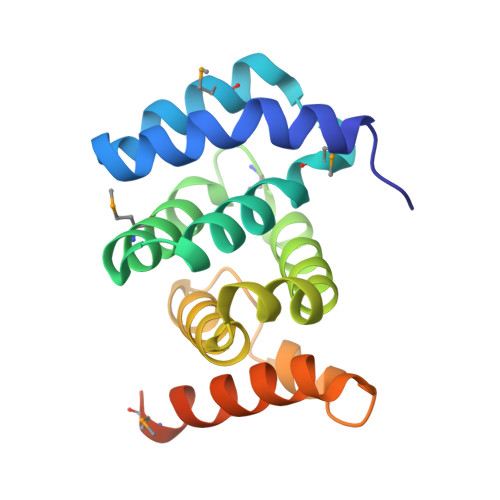
PDCD4 inhibits translation initiation by binding to eIF4A using both its MA3 domains.

Suzuki, C.Garces, R.G.Edmonds, K.A.Hiller, S.Hyberts, S.G.Marintchev, A.Wagner, G.

(2008) Proc Natl Acad Sci U S A 105: 3274-3279

  • DOI: https://doi.org/10.1073/pnas.0712235105
  • Primary Citation of Related Structures:  
    2RG8

  • PubMed Abstract: 

    Programmed Cell Death 4 (PDCD4) is a protein known to bind eukaryotic initiation factor 4A (eIF4A), inhibit translation initiation, and act as a tumor suppressor. PDCD4 contains two C-terminal MA3 domains, which are thought to be responsible for its inhibitory function. Here, we analyze the structures and inhibitory functions of these two PDCD4 MA3 domains by x-ray crystallography, NMR, and surface plasmon resonance. We show that both MA3 domains are structurally and functionally very similar and bind specifically to the eIF4A N-terminal domain (eIF4A-NTD) using similar binding interfaces. We found that the PDCD4 MA3 domains compete with the eIF4G MA3 domain and RNA for eIF4A binding. Our data provide evidence that PDCD4 inhibits translation initiation by displacing eIF4G and RNA from eIF4A. The PDCD4 MA3 domains act synergistically to form a tighter and more stable complex with eIF4A, which explains the need for two tandem MA3 domains.


  • Organizational Affiliation

    Department of Biological Chemistry and Molecular Pharmacology, Harvard Medical School, Boston, MA 02115, USA.


Macromolecules
Find similar proteins by:  (by identity cutoff)  |  3D Structure
Entity ID: 1
MoleculeChains Sequence LengthOrganismDetailsImage
Programmed cell death protein 4
A, B
165Homo sapiensMutation(s): 0 
Gene Names: PDCD4H731
UniProt & NIH Common Fund Data Resources
Find proteins for Q53EL6 (Homo sapiens)
Explore Q53EL6 
Go to UniProtKB:  Q53EL6
PHAROS:  Q53EL6
GTEx:  ENSG00000150593 
Entity Groups  
Sequence Clusters30% Identity50% Identity70% Identity90% Identity95% Identity100% Identity
UniProt GroupQ53EL6
Sequence Annotations
Expand
  • Reference Sequence
Experimental Data & Validation

Experimental Data

  • Method: X-RAY DIFFRACTION
  • Resolution: 1.80 Å
  • R-Value Free: 0.251 
  • R-Value Work: 0.202 
  • R-Value Observed: 0.202 
  • Space Group: P 21 21 21
Unit Cell:
Length ( Å )Angle ( ˚ )
a = 37.68α = 90
b = 70.06β = 90
c = 110.88γ = 90
Software Package:
Software NamePurpose
CNSrefinement
ADSCdata collection
HKL-2000data reduction
HKL-2000data scaling
SnBphasing

Structure Validation

View Full Validation Report


Entry History 

Deposition Data

Revision History  (Full details and data files)

  • Version 1.0: 2008-03-04
    Type: Initial release
  • Version 1.1: 2011-07-13
    Changes: Version format compliance
  • Version 1.2: 2017-10-25
    Changes: Refinement description