2RFN | pdb_00002rfn

x-ray structure of c-Met with inhibitor.


Experimental Data Snapshot

  • Method: X-RAY DIFFRACTION
  • Resolution: 2.50 Å
  • R-Value Free: 
    0.294 (Depositor), 0.282 (DCC) 
  • R-Value Work: 
    0.238 (Depositor), 0.231 (DCC) 
  • R-Value Observed: 
    0.241 (Depositor) 

Starting Model: experimental
View more details

wwPDB Validation 3D Report Full Report

Validation slider image for 2RFN

Ligand Structure Quality Assessment 


This is version 1.2 of the entry. See complete history

Literature

c-Met inhibitors with novel binding mode show activity against several hereditary papillary renal cell carcinoma-related mutations.

Bellon, S.F.Kaplan-Lefko, P.Yang, Y.Zhang, Y.Moriguchi, J.Rex, K.Johnson, C.W.Rose, P.E.Long, A.M.O'Connor, A.B.Gu, Y.Coxon, A.Kim, T.S.Tasker, A.Burgess, T.L.Dussault, I.

(2008) J Biological Chem 283: 2675-2683

  • DOI: https://doi.org/10.1074/jbc.M705774200
  • Primary Citation Related Structures: 
    2RFN, 2RFS

  • PubMed Abstract: 

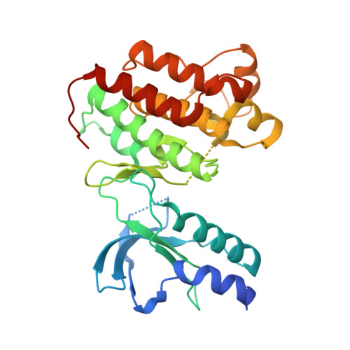
    c-Met is a receptor tyrosine kinase often deregulated in human cancers, thus making it an attractive drug target. One mechanism by which c-Met deregulation leads to cancer is through gain-of-function mutations. Therefore, small molecules capable of targeting these mutations could offer therapeutic benefits for affected patients. SU11274 was recently described and reported to inhibit the activity of the wild-type and some mutant forms of c-Met, whereas other mutants are resistant to inhibition. We identified a novel series of c-Met small molecule inhibitors that are active against multiple mutants previously identified in hereditary papillary renal cell carcinoma patients. AM7 is active against wild-type c-Met as well as several mutants, inhibits c-Met-mediated signaling in MKN-45 and U-87 MG cells, and inhibits tumor growth in these two models grown as xenografts. The crystal structures of AM7 and SU11274 bound to unphosphorylated c-Met have been determined. The AM7 structure reveals a novel binding mode compared with other published c-Met inhibitors and SU11274. The molecule binds the kinase linker and then extends into a new hydrophobic binding site. This binding site is created by a significant movement of the C-helix and so represents an inactive conformation of the c-Met kinase. Thus, our results demonstrate that it is possible to identify and design inhibitors that will likely be active against mutants found in different cancers.


  • Organizational Affiliation
    • Department of Molecular Structure, Amgen Inc., Thousand Oaks, CA 91320, USA.

Macromolecule Content 

  • Total Structure Weight: 71.92 kDa 
  • Atom Count: 4,490 
  • Modeled Residue Count: 537 
  • Deposited Residue Count: 620 
  • Unique protein chains: 1

Macromolecules

Find similar proteins by:|  3D Structure
Entity ID: 1
MoleculeChains  Sequence LengthOrganismDetailsImage
Hepatocyte growth factor receptor
A, B
310Homo sapiensMutation(s): 0 
Gene Names: MET
EC: 2.7.10.1
UniProt & NIH Common Fund Data Resources
Find proteins for P08581 (Homo sapiens)
Explore P08581 
Go to UniProtKB:  P08581
PHAROS:  P08581
GTEx:  ENSG00000105976 
Entity Groups
Sequence Clusters30% Identity50% Identity70% Identity90% Identity95% Identity100% Identity
UniProt GroupP08581
Sequence Annotations
Expand
Reference Sequence

Small Molecules

Ligands 1 Unique
IDChains Name / Formula / InChI Key2D Diagram3D Interactions
AM7

Query on AM7


Download:Ideal Coordinates CCD File
C [auth A],
D [auth B]
2-benzyl-5-(3-fluoro-4-{[6-methoxy-7-(3-morpholin-4-ylpropoxy)quinolin-4-yl]oxy}phenyl)-3-methylpyrimidin-4(3H)-one
C35 H35 F N4 O5
PEGWVOKOYYAQEV-UHFFFAOYSA-N
Binding Affinity Annotations 
IDSourceBinding Affinity
AM7 BindingDB:  2RFN Ki: 8.9 (nM) from 1 assay(s)
IC50: min: 8.9, max: 17 (nM) from 2 assay(s)

Experimental Data & Validation

Experimental Data

  • Method: X-RAY DIFFRACTION
  • Resolution: 2.50 Å
  • R-Value Free:  0.294 (Depositor), 0.282 (DCC) 
  • R-Value Work:  0.238 (Depositor), 0.231 (DCC) 
  • R-Value Observed: 0.241 (Depositor) 
Space Group: P 21 21 21
Unit Cell:
Length ( Å )Angle ( ˚ )
a = 74.509α = 90
b = 78.919β = 90
c = 129.128γ = 90
Software Package:
Software NamePurpose
REFMACrefinement
CrystalCleardata collection
DENZOdata reduction
SCALEPACKdata scaling
AMoREphasing

Structure Validation

View Full Validation Report


Ligand Structure Quality Assessment 


Entry History 

Deposition Data

Revision History  (Full details and data files)

  • Version 1.0: 2007-11-06
    Type: Initial release
  • Version 1.1: 2011-07-13
    Changes: Advisory, Version format compliance
  • Version 1.2: 2023-08-30
    Changes: Data collection, Database references, Derived calculations, Refinement description