2RCB

Crystal structure of the NR3B ligand binding core complex with D-serine at 1.62 Angstrom resolution


Experimental Data Snapshot

  • Method: X-RAY DIFFRACTION
  • Resolution: 1.62 Å
  • R-Value Free: 0.228 
  • R-Value Work: 0.192 
  • R-Value Observed: 0.195 

wwPDB Validation   3D Report Full Report


This is version 1.3 of the entry. See complete history


Literature

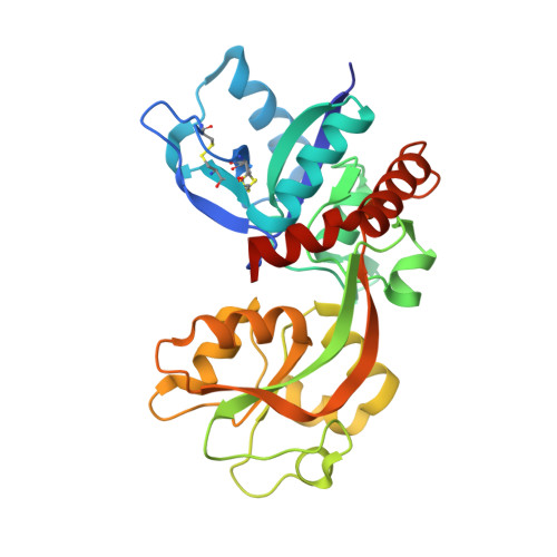
Molecular mechanism of ligand recognition by NR3 subtype glutamate receptors.

Yao, Y.Harrison, C.B.Freddolino, P.L.Schulten, K.Mayer, M.L.

(2008) EMBO J 27: 2158-2170

  • DOI: https://doi.org/10.1038/emboj.2008.140
  • Primary Citation of Related Structures:  
    2RC7, 2RC8, 2RC9, 2RCA, 2RCB

  • PubMed Abstract: 

    NR3 subtype glutamate receptors have a unique developmental expression profile, but are the least well-characterized members of the NMDA receptor gene family, which have key roles in synaptic plasticity and brain development. Using ligand binding assays, crystallographic analysis, and all atom MD simulations, we investigate mechanisms underlying the binding by NR3A and NR3B of glycine and D-serine, which are candidate neurotransmitters for NMDA receptors containing NR3 subunits. The ligand binding domains of both NR3 subunits adopt a similar extent of domain closure as found in the corresponding NR1 complexes, but have a unique loop 1 structure distinct from that in all other glutamate receptor ion channels. Within their ligand binding pockets, NR3A and NR3B have strikingly different hydrogen bonding networks and solvent structures from those found in NR1, and fail to undergo a conformational rearrangement observed in NR1 upon binding the partial agonist ACPC. MD simulations revealed numerous interdomain contacts, which stabilize the agonist-bound closed-cleft conformation, and a novel twisting motion for the loop 1 helix that is unique in NR3 subunits.


  • Organizational Affiliation

    Laboratory of Cellular and Molecular Neurophysiology, Porter Neuroscience Research Center, NICHD, NIH, DHHS, Bethesda, MD, USA.


Macromolecules
Find similar proteins by:  (by identity cutoff)  |  3D Structure
Entity ID: 1
MoleculeChains Sequence LengthOrganismDetailsImage
Glutamate [NMDA] receptor subunit 3B
A, B
292Rattus norvegicusMutation(s): 0 
Gene Names: Grin3b
UniProt
Find proteins for Q8VHN2 (Rattus norvegicus)
Explore Q8VHN2 
Go to UniProtKB:  Q8VHN2
Entity Groups  
Sequence Clusters30% Identity50% Identity70% Identity90% Identity95% Identity100% Identity
UniProt GroupQ8VHN2
Sequence Annotations
Expand
  • Reference Sequence
Small Molecules
Binding Affinity Annotations 
IDSourceBinding Affinity
DSN PDBBind:  2RCB Kd: 163 (nM) from 1 assay(s)
Experimental Data & Validation

Experimental Data

  • Method: X-RAY DIFFRACTION
  • Resolution: 1.62 Å
  • R-Value Free: 0.228 
  • R-Value Work: 0.192 
  • R-Value Observed: 0.195 
  • Space Group: P 21 21 21
Unit Cell:
Length ( Å )Angle ( ˚ )
a = 45.779α = 90
b = 83.521β = 90
c = 144.959γ = 90
Software Package:
Software NamePurpose
REFMACrefinement
HKL-2000data collection
HKL-2000data reduction
HKL-2000data scaling
PHASERphasing

Structure Validation

View Full Validation Report


Entry History 

Deposition Data

Revision History  (Full details and data files)

  • Version 1.0: 2008-08-05
    Type: Initial release
  • Version 1.1: 2011-07-13
    Changes: Advisory, Version format compliance
  • Version 1.2: 2017-08-23
    Changes: Source and taxonomy
  • Version 1.3: 2023-08-30
    Changes: Data collection, Database references, Derived calculations, Refinement description