2R5Y | pdb_00002r5y

Structure of Scr/Exd complex bound to a consensus Hox-Exd site


Experimental Data Snapshot

  • Method: X-RAY DIFFRACTION
  • Resolution: 2.60 Å
  • R-Value Free: 
    0.299 (Depositor), 0.302 (DCC) 
  • R-Value Work: 
    0.255 (Depositor), 0.254 (DCC) 
  • R-Value Observed: 
    0.255 (Depositor) 

Starting Model: experimental
View more details

wwPDB Validation 3D Report Full Report

Validation slider image for 2R5Y

This is version 1.3 of the entry. See complete history

Literature

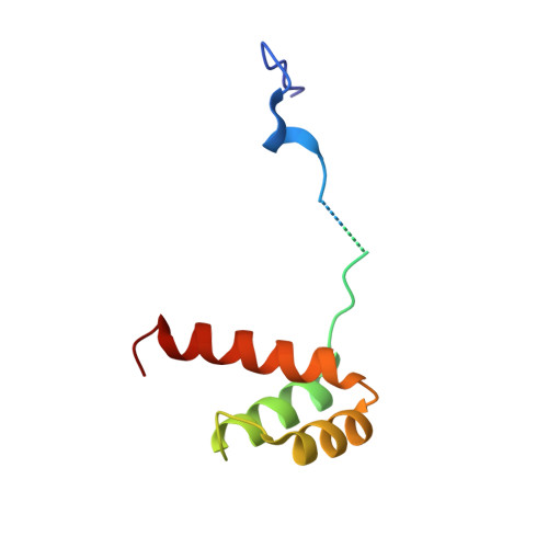
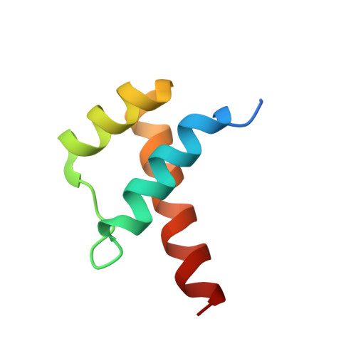
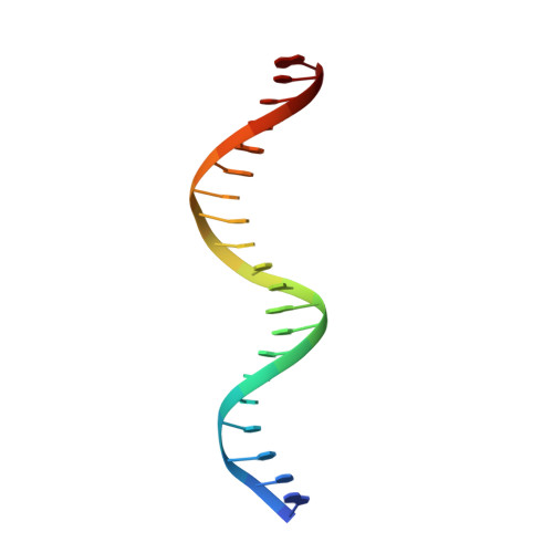
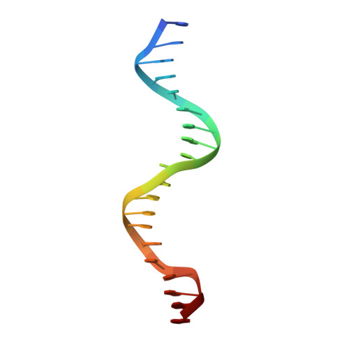
Functional specificity of a Hox protein mediated by the recognition of minor groove structure

Joshi, R.Passner, J.M.Rohs, R.Jain, R.Sosinsky, A.Crickmore, M.A.Jacob, V.Aggarwal, A.K.Honig, B.Mann, R.S.

(2007) Cell 131: 530-543

  • DOI: https://doi.org/10.1016/j.cell.2007.09.024
  • Primary Citation Related Structures: 
    2R5Y, 2R5Z

  • PubMed Abstract: 

    The recognition of specific DNA-binding sites by transcription factors is a critical yet poorly understood step in the control of gene expression. Members of the Hox family of transcription factors bind DNA by making nearly identical major groove contacts via the recognition helices of their homeodomains. In vivo specificity, however, often depends on extended and unstructured regions that link Hox homeodomains to a DNA-bound cofactor, Extradenticle (Exd). Using a combination of structure determination, computational analysis, and in vitro and in vivo assays, we show that Hox proteins recognize specific Hox-Exd binding sites via residues located in these extended regions that insert into the minor groove but only when presented with the correct DNA sequence. Our results suggest that these residues, which are conserved in a paralog-specific manner, confer specificity by recognizing a sequence-dependent DNA structure instead of directly reading a specific DNA sequence.


  • Organizational Affiliation
    • Department of Biochemistry and Molecular Biophysics, Columbia University, HHSC 1104, New York, NY 10032, USA.

Macromolecule Content 

  • Total Structure Weight: 30.79 kDa 
  • Atom Count: 2,066 
  • Modeled Residue Count: 172 
  • Deposited Residue Count: 191 
  • Unique protein chains: 2
  • Unique nucleic acid chains: 2

Macromolecules


Find similar proteins by:|  3D Structure
Entity ID: 3
MoleculeChains  Sequence LengthOrganismDetailsImage
Homeotic protein Sex combs reducedC [auth A]88Drosophila melanogasterMutation(s): 1 
Gene Names: Scr
UniProt
Find proteins for P09077 (Drosophila melanogaster)
Explore P09077 
Go to UniProtKB:  P09077
Entity Groups
Sequence Clusters30% Identity50% Identity70% Identity90% Identity95% Identity100% Identity
UniProt GroupP09077
Sequence Annotations
Expand
Reference Sequence
Find similar proteins by:|  3D Structure
Entity ID: 4
MoleculeChains  Sequence LengthOrganismDetailsImage
Homeobox protein extradenticleD [auth B]63Drosophila melanogasterMutation(s): 0 
Gene Names: exd
UniProt
Find proteins for P40427 (Drosophila melanogaster)
Explore P40427 
Go to UniProtKB:  P40427
Entity Groups
Sequence Clusters30% Identity50% Identity70% Identity90% Identity95% Identity100% Identity
UniProt GroupP40427
Sequence Annotations
Expand
Reference Sequence
Find similar nucleic acids by:  Sequence
Entity ID: 1
MoleculeChains LengthOrganismImage
DNA (5'-D(*DAP*DCP*DTP*DCP*DTP*DAP*DTP*DGP*DAP*DTP*DTP*DTP*DAP*DTP*DGP*DGP*DGP*DCP*DTP*DG)-3')A [auth C]20synthetic construct
Sequence Annotations
Expand
Reference Sequence
Find similar nucleic acids by:  Sequence
Entity ID: 2
MoleculeChains LengthOrganismImage
DNA (5'-D(*DTP*DCP*DAP*DGP*DCP*DCP*DCP*DAP*DTP*DAP*DAP*DAP*DTP*DCP*DAP*DTP*DAP*DGP*DAP*DG)-3')B [auth D]20synthetic construct
Sequence Annotations
Expand
Reference Sequence

Experimental Data & Validation

Experimental Data

  • Method: X-RAY DIFFRACTION
  • Resolution: 2.60 Å
  • R-Value Free:  0.299 (Depositor), 0.302 (DCC) 
  • R-Value Work:  0.255 (Depositor), 0.254 (DCC) 
  • R-Value Observed: 0.255 (Depositor) 
Space Group: P 43 21 2
Unit Cell:
Length ( Å )Angle ( ˚ )
a = 65.31α = 90
b = 65.31β = 90
c = 200.3γ = 90
Software Package:
Software NamePurpose
CNSrefinement
HKL-2000data collection
HKL-2000data reduction
HKL-2000data scaling
AMoREphasing

Structure Validation

View Full Validation Report


Entry History 

Deposition Data

Revision History  (Full details and data files)

  • Version 1.0: 2008-02-05
    Type: Initial release
  • Version 1.1: 2011-07-13
    Changes: Version format compliance
  • Version 1.2: 2021-10-20
    Changes: Database references, Source and taxonomy
  • Version 1.3: 2023-08-30
    Changes: Data collection, Refinement description