Mutational and structural studies of the active-site residues in truncated Fibrobacter succinogenes1,3-1,4-beta-D-glucanase.
Tsai, L.C., Huang, H.C., Hsiao, C.H., Chiang, Y.N., Shyur, L.F., Lin, Y.S., Lee, S.H.(2008) Acta Crystallogr D Biol Crystallogr 64: 1259-1266
- PubMed: 19018102 Search on PubMed
- DOI: https://doi.org/10.1107/S0907444908033428
- Primary Citation Related Structures:
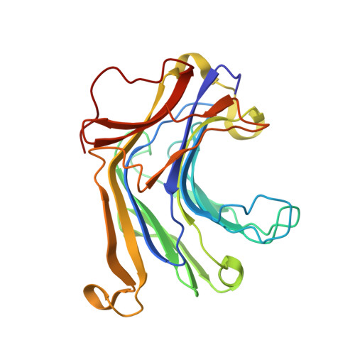
2R49 - PubMed Abstract:
1,3-1,4-beta-D-Glucanases (EC 3.2.1.73) specifically hydrolyze beta-1,4-glycosidic bonds located prior to beta-1,3-glycosidic linkages in lichenan or beta-D-glucans. It has been suggested that truncated Fibrobacter succinogenes 1,3-1,4-beta-D-glucanase (TFsbeta-glucanase) can accommodate five glucose rings in its active site upon enzyme-substrate interaction. In this study, 12 mutant enzymes were created by mutating the conserved residues Gln70, Asn72, Gln81 and Glu85 proposed to bind to substrate subsites +1 and +2 and the catalytic properties of these mutants were determined. The most significant change in catalytic activity was observed on mutation of Gln70, with a 299-fold and 498-fold lower k(cat)/K(m) for the mutants Q70A and Q70I, respectively, compared with the wild-type enzyme. Mutagenesis, kinetic and structural studies revealed that the conserved residues surrounding the active site of TFsbeta-glucanase at substrate subsites +1 and +2 play an important role in its catalytic function, with the following order of importance: Gln70 > Asn72 > Glu85 > Gln81. The crystal structure of mutant E85I was determined at 2.2 A resolution. Further analysis of the E85I mutant structure revealed that the loop located at the concave site moved approximately 2 A from its position in the native enzyme complex without changing the core structure.
- Department of Molecular Science and Engineering, National Taipei University of Technology, Taipei 106, Taiwan. lichu@ntut.edu.tw
Organizational Affiliation: