2QZF

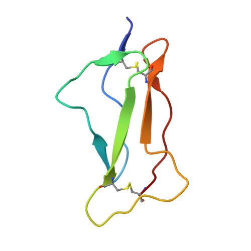
SCR1 of DAF from 1ojv fitted into cryoEM density


Experimental Data Snapshot

  • Method: ELECTRON MICROSCOPY
  • Resolution: 14.0 Å
  • Aggregation State: PARTICLE 
  • Reconstruction Method: SINGLE PARTICLE 

wwPDB Validation   3D Report Full Report


This is version 1.3 of the entry. See complete history


Literature

Interaction of decay-accelerating factor with coxsackievirus b3.

Hafenstein, S.Bowman, V.D.Chipman, P.R.Kelly, C.M.Lin, F.Medof, M.E.Rossmann, M.G.

(2007) J Virol 81: 12927-12935

  • DOI: https://doi.org/10.1128/JVI.00931-07
  • Primary Citation of Related Structures:  
    2QZD, 2QZF, 2QZH

  • PubMed Abstract: 

    Many entero-, parecho-, and rhinoviruses use immunoglobulin (Ig)-like receptors that bind into the viral canyon and are required to initiate viral uncoating during infection. However, some of these viruses use an alternative or additional receptor that binds outside the canyon. Both the coxsackievirus-adenovirus receptor (CAR), an Ig-like molecule that binds into the viral canyon, and decay-accelerating factor (DAF) have been identified as cellular receptors for coxsackievirus B3 (CVB3). A cryoelectron microscopy reconstruction of a variant of CVB3 complexed with DAF shows full occupancy of the DAF receptor in each of 60 binding sites. The DAF molecule bridges the canyon, blocking the CAR binding site and causing the two receptors to compete with one another. The binding site of DAF on CVB3 differs from the binding site of DAF on the surface of echoviruses, suggesting independent evolutionary processes.


  • Organizational Affiliation

    Department of Biological Sciences, Purdue University, 915 W. State Street, West Lafayette, IN 47907-2054, USA.


Macromolecules
Find similar proteins by:  (by identity cutoff)  |  3D Structure
Entity ID: 1
MoleculeChains Sequence LengthOrganismDetailsImage
Complement decay-accelerating factor62Homo sapiensMutation(s): 0 
UniProt & NIH Common Fund Data Resources
Find proteins for P08174 (Homo sapiens)
Explore P08174 
Go to UniProtKB:  P08174
PHAROS:  P08174
GTEx:  ENSG00000196352 
Entity Groups  
Sequence Clusters30% Identity50% Identity70% Identity90% Identity95% Identity100% Identity
UniProt GroupP08174
Sequence Annotations
Expand
  • Reference Sequence
Experimental Data & Validation

Experimental Data

  • Method: ELECTRON MICROSCOPY
  • Resolution: 14.0 Å
  • Aggregation State: PARTICLE 
  • Reconstruction Method: SINGLE PARTICLE 
EM Software:
TaskSoftware PackageVersion
RECONSTRUCTIONEM3DR
RECONSTRUCTIONEMPFT

Structure Validation

View Full Validation Report


Entry History 

Deposition Data

Revision History  (Full details and data files)

  • Version 1.0: 2007-10-30
    Type: Initial release
  • Version 1.1: 2011-07-13
    Changes: Version format compliance
  • Version 1.2: 2012-05-30
    Changes: Refinement description
  • Version 1.3: 2018-07-18
    Changes: Data collection