Determining Rieske cluster reduction potentials.
Brown, E.N., Friemann, R., Karlsson, A., Parales, J.V., Couture, M.M., Eltis, L.D., Ramaswamy, S.(2008) J Biol Inorg Chem 13: 1301-1313
- PubMed: 18719951 Search on PubMed
- DOI: https://doi.org/10.1007/s00775-008-0413-4
- Primary Citation Related Structures:
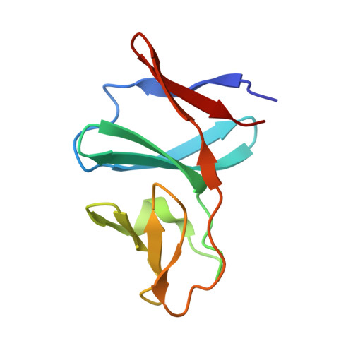
2QPZ - PubMed Abstract:
The Rieske iron-sulfur proteins have reduction potentials ranging from -150 to +400 mV. This enormous range of potentials was first proposed to be due to differing solvent exposure or even protein structure. However, the increasing number of available crystal structures for Rieske iron-sulfur proteins has shown this not to be the case. Colbert and colleagues proposed in 2000 that differences in the electrostatic environment, and not structural differences, of a Rieske proteins are responsible for the wide range of reduction potentials observed. Using computational simulation methods and the newly determined structure of Pseudomonas sp. NCIB 9816-4 naphthalene dioxygenase Rieske ferredoxin (NDO-F9816-4), we have developed a model to predict the reduction potential of Rieske proteins given only their crystal structure. The reduction potential of NDO-F9816-4, determined using a highly oriented pyrolytic graphite electrode, was -150+/-2 mV versus the standard hydrogen electrode. The predicted reduction potentials correlate well with experimentally determined potentials. Given this model, the effect of protein mutations can be evaluated. Our results suggest that the reduction potential of new proteins can be estimated with good confidence from 3D structures of proteins. The structure of NDO-F9816-4 is the most basic Rieske ferredoxin structure determined to date. Thus, the contributions of additional structural motifs and their effects on reduction potential can be compared with respect to this base structure.
- Department of Biochemistry, University of Iowa, Iowa City, IA 52242, USA.
Organizational Affiliation: