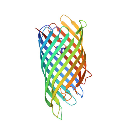
2QOM

The crystal structure of the E.coli EspP autotransporter Beta-domain.


Experimental Data Snapshot

  • Method: X-RAY DIFFRACTION
  • Resolution: 2.66 Å
  • R-Value Free: 0.284 
  • R-Value Work: 0.258 
  • R-Value Observed: 0.260 

Starting Model: experimental
View more details

wwPDB Validation   3D Report Full Report


This is version 1.4 of the entry. See complete history


Literature

Autotransporter structure reveals intra-barrel cleavage followed by conformational changes.

Barnard, T.J.Dautin, N.Lukacik, P.Bernstein, H.D.Buchanan, S.K.

(2007) Nat Struct Mol Biol 14: 1214-1220

  • DOI: https://doi.org/10.1038/nsmb1322
  • Primary Citation of Related Structures:  
    2QOM

  • PubMed Abstract: 

    Autotransporters are virulence factors produced by Gram-negative bacteria. They consist of two domains, an N-terminal 'passenger' domain and a C-terminal beta-domain. beta-domains form beta-barrel structures in the outer membrane while passenger domains are translocated into the extracellular space. In some autotransporters, the two domains are separated by proteolytic cleavage. Using X-ray crystallography, we solved the 2.7-A structure of the post-cleavage state of the beta-domain of EspP, an autotransporter produced by Escherichia coli strain O157:H7. The structure consists of a 12-stranded beta-barrel with the passenger domain-beta-domain cleavage junction located inside the barrel pore, approximately midway between the extracellular and periplasmic surfaces of the outer membrane. The structure reveals an unprecedented intra-barrel cleavage mechanism and suggests that two conformational changes occur in the beta-domain after cleavage, one conferring increased stability on the beta-domain and another restricting access to the barrel pore.


  • Organizational Affiliation

    Laboratory of Molecular Biology, National Institute of Diabetes and Digestive and Kidney Diseases, US National Institutes of Health, Bethesda, Maryland 20892, USA.


Macromolecules
Find similar proteins by:  (by identity cutoff)  |  3D Structure
Entity ID: 1
MoleculeChains Sequence LengthOrganismDetailsImage
Serine protease espP
A, B
285Escherichia coli O157:H7Mutation(s): 0 
Gene Names: espP
EC: 3.4.21
Membrane Entity: Yes 
UniProt
Find proteins for Q7BSW5 (Escherichia coli O157:H7)
Explore Q7BSW5 
Go to UniProtKB:  Q7BSW5
Entity Groups  
Sequence Clusters30% Identity50% Identity70% Identity90% Identity95% Identity100% Identity
UniProt GroupQ7BSW5
Sequence Annotations
Expand
  • Reference Sequence
Experimental Data & Validation

Experimental Data

  • Method: X-RAY DIFFRACTION
  • Resolution: 2.66 Å
  • R-Value Free: 0.284 
  • R-Value Work: 0.258 
  • R-Value Observed: 0.260 
  • Space Group: P 1 21 1
Unit Cell:
Length ( Å )Angle ( ˚ )
a = 84.951α = 90
b = 53.298β = 103.57
c = 102.418γ = 90
Software Package:
Software NamePurpose
REFMACrefinement
HKL-2000data reduction
HKL-2000data scaling
PHENIXphasing

Structure Validation

View Full Validation Report


Entry History 

Deposition Data

Revision History  (Full details and data files)

  • Version 1.0: 2007-11-13
    Type: Initial release
  • Version 1.1: 2011-07-13
    Changes: Advisory, Derived calculations, Source and taxonomy, Version format compliance
  • Version 1.2: 2017-10-25
    Changes: Refinement description
  • Version 1.3: 2024-02-21
    Changes: Data collection, Database references, Refinement description
  • Version 1.4: 2024-04-03
    Changes: Refinement description