Identification of CD46 binding sites within the adenovirus serotype 35 fiber knob
Wang, H., Liaw, Y.C., Stone, D., Kalyuzhniy, O., Amiraslanov, I., Tuve, S., Verlinde, C.L., Shayakhmetov, D., Stehle, T., Roffler, S., Lieber, A.(2007) J Virol 81: 12785-12792
- PubMed: 17898059 Search on PubMedSearch on PubMed Central
- DOI: https://doi.org/10.1128/JVI.01732-07
- Primary Citation Related Structures:
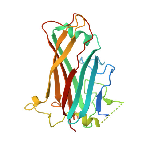
2QLK - PubMed Abstract:
Species B human adenoviruses (Ads) are often associated with fatal illnesses in immunocompromised individuals. Recently, species B Ads, most of which use the ubiquitously expressed complement regulatory protein CD46 as a primary attachment receptor, have gained interest for use as gene therapy vectors. In this study, we focused on species B Ad serotype 35 (Ad35), whose trimeric fiber knob domain binds to three CD46 molecules with a KD (equilibrium dissociation constant) of 15.5 nM. To study the Ad35 knob-CD46 interaction, we generated an expression library of Ad35 knobs with random mutations and screened it for CD46 binding. We identified four critical residues (Phe242, Arg279, Ser282, and Glu302) which, when mutated, ablated Ad35 knob binding to CD46 without affecting knob trimerization. The functional importance of the identified residues was validated in surface plasmon resonance and competition binding studies. To model the Ad35 knob-CD46 interaction, we resolved the Ad35 knob structure at 2-A resolution by X-ray crystallography and overlaid it onto the existing structure for Ad11-CD46 interaction. According to our model, all identified Ad35 residues are in regions that interact with CD46, whereby one CD46 molecule binds between two knob monomers. This mode of interaction might have potential consequences for CD46 signaling and intracellular trafficking of Ad35. Our findings are also fundamental for better characterization of species B Ads and design of antiviral drugs, as well as for application of species B Ads as in vivo and in vitro gene transfer vectors.
- Division of Medical Genetics, University of Washington, Box 357720, Seattle, WA 98195, USA.
Organizational Affiliation: