2QD4 | pdb_00002qd4

Wild type human ferrochelatase crystallized with MnCl2


Experimental Data Snapshot

  • Method: X-RAY DIFFRACTION
  • Resolution: 2.00 Å
  • R-Value Free: 
    0.230 (Depositor), 0.225 (DCC) 
  • R-Value Work: 
    0.206 (Depositor), 0.202 (DCC) 
  • R-Value Observed: 
    0.206 (Depositor) 

wwPDB Validation 3D Report Full Report

Validation slider image for 2QD4

Ligand Structure Quality Assessment 


This is version 1.2 of the entry. See complete history

Literature

A pi-Helix Switch Selective for Porphyrin Deprotonation and Product Release in Human Ferrochelatase.

Medlock, A.E.Dailey, T.A.Ross, T.A.Dailey, H.A.Lanzilotta, W.N.

(2007) J Mol Biology 373: 1006-1016

  • DOI: https://doi.org/10.1016/j.jmb.2007.08.040
  • Primary Citation Related Structures: 
    2QD1, 2QD2, 2QD3, 2QD4, 2QD5

  • PubMed Abstract: 

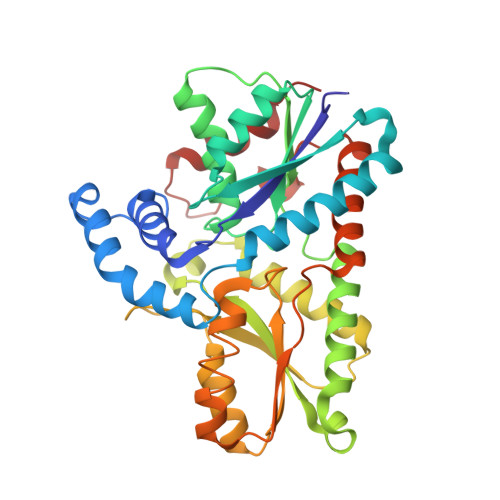
    Ferrochelatase (protoheme ferrolyase, EC 4.99.1.1) is the terminal enzyme in heme biosynthesis and catalyzes the insertion of ferrous iron into protoporphyrin IX to form protoheme IX (heme). Due to the many critical roles of heme, synthesis of heme is required by the vast majority of organisms. Despite significant investigation of both the microbial and eukaryotic enzyme, details of metal chelation remain unidentified. Here we present the first structure of the wild-type human enzyme, a lead-inhibited intermediate of the wild-type enzyme with bound metallated porphyrin macrocycle, the product bound form of the enzyme, and a higher resolution model for the substrate-bound form of the E343K variant. These data paint a picture of an enzyme that undergoes significant changes in secondary structure during the catalytic cycle. The role that these structural alterations play in overall catalysis and potential protein-protein interactions with other proteins, as well as the possible molecular basis for these changes, is discussed. The atomic details and structural rearrangements presented herein significantly advance our understanding of the substrate binding mode of ferrochelatase and reveal new conformational changes in a structurally conserved pi-helix that is predicted to have a central role in product release.


  • Organizational Affiliation
    • Biomedical and Health Sciences Institute, Department of Biochemistry and Molecular Biology, University of Georgia, Athens, GA 30602, USA.

Macromolecule Content 

  • Total Structure Weight: 85.46 kDa 
  • Atom Count: 6,580 
  • Modeled Residue Count: 718 
  • Deposited Residue Count: 718 
  • Unique protein chains: 1

Macromolecules

Find similar proteins by:|  3D Structure
Entity ID: 1
MoleculeChains  Sequence LengthOrganismDetailsImage
Ferrochelatase
A, B
359Homo sapiensMutation(s): 0 
Gene Names: FECH
EC: 4.99.1.1 (PDB Primary Data), 4.98.1.1 (UniProt)
UniProt & NIH Common Fund Data Resources
Find proteins for P22830 (Homo sapiens)
Explore P22830 
Go to UniProtKB:  P22830
PHAROS:  P22830
GTEx:  ENSG00000066926 
Entity Groups
Sequence Clusters30% Identity50% Identity70% Identity90% Identity95% Identity100% Identity
UniProt GroupP22830
Sequence Annotations
Expand
Reference Sequence

Small Molecules

Ligands 4 Unique
IDChains Name / Formula / InChI Key2D Diagram3D Interactions
CHD

Query on CHD


Download:Ideal Coordinates CCD File
H [auth A]
I [auth A]
J [auth A]
O [auth B]
P [auth B]
H [auth A],
I [auth A],
J [auth A],
O [auth B],
P [auth B],
Q [auth B]
CHOLIC ACID
C24 H40 O5
BHQCQFFYRZLCQQ-OELDTZBJSA-N
FES

Query on FES


Download:Ideal Coordinates CCD File
G [auth A],
N [auth B]
FE2/S2 (INORGANIC) CLUSTER
Fe2 S2
NIXDOXVAJZFRNF-UHFFFAOYSA-N
GOL

Query on GOL


Download:Ideal Coordinates CCD File
K [auth A]GLYCEROL
C3 H8 O3
PEDCQBHIVMGVHV-UHFFFAOYSA-N
CL

Query on CL


Download:Ideal Coordinates CCD File
C [auth A]
D [auth A]
E [auth A]
F [auth A]
L [auth B]
C [auth A],
D [auth A],
E [auth A],
F [auth A],
L [auth B],
M [auth B]
CHLORIDE ION
Cl
VEXZGXHMUGYJMC-UHFFFAOYSA-M

Experimental Data & Validation

Experimental Data

  • Method: X-RAY DIFFRACTION
  • Resolution: 2.00 Å
  • R-Value Free:  0.230 (Depositor), 0.225 (DCC) 
  • R-Value Work:  0.206 (Depositor), 0.202 (DCC) 
  • R-Value Observed: 0.206 (Depositor) 
Space Group: P 21 21 21
Unit Cell:
Length ( Å )Angle ( ˚ )
a = 87.586α = 90
b = 93.062β = 90
c = 109.98γ = 90
Software Package:
Software NamePurpose
CNSrefinement
HKL-2000data collection
HKL-2000data reduction
HKL-2000data scaling
CNSphasing

Structure Validation

View Full Validation Report


Ligand Structure Quality Assessment 


Entry History 

Deposition Data

Revision History  (Full details and data files)

  • Version 1.0: 2007-10-30
    Type: Initial release
  • Version 1.1: 2011-07-13
    Changes: Derived calculations, Version format compliance
  • Version 1.2: 2024-02-21
    Changes: Data collection, Database references, Derived calculations