Structural Studies of the Final Enzyme in the alpha-Aminoadipate Pathway-Saccharopine Dehydrogenase from Saccharomyces cerevisiae
Burk, D.L., Hwang, J., Kwok, E., Marrone, L., Goodfellow, V., Dmitrienko, G.I., Berghuis, A.M.(2007) J Mol Biology 373: 745-754
- PubMed: 17854830 Search on PubMed
- DOI: https://doi.org/10.1016/j.jmb.2007.08.044
- Primary Citation Related Structures:
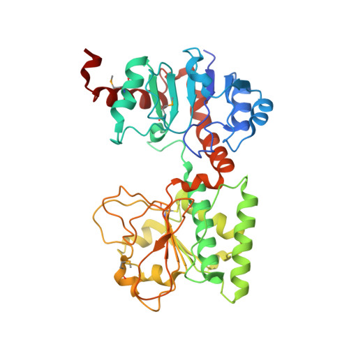
2Q99 - PubMed Abstract:
The 1.64 A structure of the apoenzyme form of saccharopine dehydrogenase (SDH) from Saccharomyces cerevisiae shows the enzyme to be composed of two domains with similar dinucleotide binding folds with a deep cleft at the interface. The structure reveals homology to alanine dehydrogenase, despite low primary sequence similarity. A model of the ternary complex of SDH, NAD, and saccharopine identifies residues Lys77 and Glu122 as potentially important for substrate binding and/or catalysis, consistent with a proton shuttle mechanism. Furthermore, the model suggests that a conformational change is required for catalysis and that residues Lys99 and Asp281 may be instrumental in mediating this change. Analysis of the crystal structure in the context of other homologous enzymes from pathogenic fungi and human sources sheds light into the suitability of SDH as a target for antimicrobial drug development.
- Department of Biochemistry, McGill University, Montreal, Quebec, Canada H3A 1A4.
Organizational Affiliation: