


The Crystallographic Structure of Alr2-Idd393 Complex Confirms Leu300 as a Specificity Determinant
Ruiz, F., Hazemann, I., Darmanin, C., Mitschler, A., Van Zandt, M., Joachimiak, A., El-Kabbani, O., Podjarny, A.To be published.
Experimental Data Snapshot
Starting Model: experimental
View more details
Entity ID: 1 | |||||
|---|---|---|---|---|---|
| Molecule | Chains | Sequence Length | Organism | Details | Image |
| Aldose reductase | 316 | Homo sapiens | Mutation(s): 0 Gene Names: ALDR1 EC: 1.1.1.21 (PDB Primary Data), 1.1.1.372 (UniProt), 1.1.1.300 (UniProt), 1.1.1.54 (UniProt) | 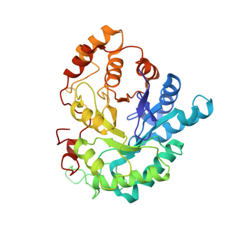 | |
UniProt & NIH Common Fund Data Resources | |||||
PHAROS: P15121 GTEx: ENSG00000085662 | |||||
Entity Groups | |||||
| Sequence Clusters | 30% Identity50% Identity70% Identity90% Identity95% Identity100% Identity | ||||
| UniProt Group | P15121 | ||||
Sequence AnnotationsExpand | |||||
Reference Sequence | |||||
| Ligands 2 Unique | |||||
|---|---|---|---|---|---|
| ID | Chains | Name / Formula / InChI Key | 2D Diagram | 3D Interactions | |
| NAP Download:Ideal Coordinates CCD File | B [auth A] | NADP NICOTINAMIDE-ADENINE-DINUCLEOTIDE PHOSPHATE C21 H28 N7 O17 P3 XJLXINKUBYWONI-NNYOXOHSSA-N |  | ||
| 393 Download:Ideal Coordinates CCD File | C [auth A] | (5-CHLORO-2-{[(3-NITROBENZYL)AMINO]CARBONYL}PHENOXY)ACETIC ACID C16 H13 Cl N2 O6 VABIMMIJVWNHFI-UHFFFAOYSA-N |  | ||
| Length ( Å ) | Angle ( ˚ ) |
|---|---|
| a = 47.224 | α = 67.58 |
| b = 47.154 | β = 76.47 |
| c = 40.316 | γ = 76.11 |
| Software Name | Purpose |
|---|---|
| SHELX | model building |
| SHELXL-97 | refinement |
| Propietary | data collection |
| HKL-2000 | data reduction |
| SCALEPACK | data scaling |
| AMoRE | phasing |


