2PSP

Porcine pancreatic spasmolytic polypeptide


Experimental Data Snapshot

  • Method: X-RAY DIFFRACTION
  • Resolution: 1.95 Å
  • R-Value Free: 0.258 
  • R-Value Work: 0.198 
  • R-Value Observed: 0.198 

wwPDB Validation   3D Report Full Report


This is version 2.0 of the entry. See complete history


Literature

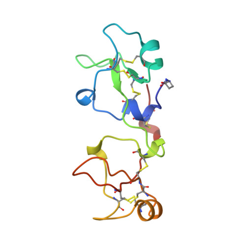
Structure of porcine pancreatic spasmolytic polypeptide at 1.95 A resolution.

Petersen, T.N.Henriksen, A.Gajhede, M.

(1996) Acta Crystallogr D Biol Crystallogr 52: 730-737

  • DOI: https://doi.org/10.1107/S0907444996001345
  • Primary Citation of Related Structures:  
    2PSP

  • PubMed Abstract: 

    The structure of a trigonal crystal form of porcine pancreatic spasmolytic polypeptide (PSP) has been solved by molecular replacement and refined to 1.95 A resolution. Three heavy-atom derivatives were prepared, giving unbiased phase information, which was used in the model building of the protein molecules. The final conventional R value is 19.8% with the inclusion of 183 water molecules. PSP crystallizes as a dimer in space group P3(1)21 with a non-crystallographic twofold axis relating the monomers. The monomer consists of two very similar domains each composed of three loop regions. Two clefts are found in the monomer, one in each domain, that are proposed as possible substrate-binding sites. Important interactions have been identified in the proposed substrate-binding sites, where conserved water molecules probably mimic the hydrophilic positions of the substrate. The estimated cleft size is 9 x 9 x 12 A. Analysis of the charge distribution within the clefts, by an electrostatic potential calculation, shows the clefts to be essentially non-charged.


  • Organizational Affiliation

    Centre for Crystallographic Studies, Universitetsparken, Department of Chemsistry, University of Copenhagen, Denmark.


Macromolecules
Find similar proteins by:  (by identity cutoff)  |  3D Structure
Entity ID: 1
MoleculeChains Sequence LengthOrganismDetailsImage
PORCINE PANCREATIC SPASMOLYTIC POLYPEPTIDE
A, B
106Sus scrofaMutation(s): 0 
UniProt
Find proteins for P01359 (Sus scrofa)
Explore P01359 
Go to UniProtKB:  P01359
Entity Groups  
Sequence Clusters30% Identity50% Identity70% Identity90% Identity95% Identity100% Identity
UniProt GroupP01359
Sequence Annotations
Expand
  • Reference Sequence
Small Molecules
Modified Residues  1 Unique
IDChains TypeFormula2D DiagramParent
PCA
Query on PCA
A, B
L-PEPTIDE LINKINGC5 H7 N O3GLN
Experimental Data & Validation

Experimental Data

  • Method: X-RAY DIFFRACTION
  • Resolution: 1.95 Å
  • R-Value Free: 0.258 
  • R-Value Work: 0.198 
  • R-Value Observed: 0.198 
  • Space Group: P 31 2 1
Unit Cell:
Length ( Å )Angle ( ˚ )
a = 60.6α = 90
b = 60.6β = 90
c = 113.1γ = 120
Software Package:
Software NamePurpose
DENZOdata reduction
X-PLORmodel building
X-PLORrefinement
X-PLORphasing

Structure Validation

View Full Validation Report


Entry History 

Deposition Data

Revision History  (Full details and data files)

  • Version 1.0: 1996-07-11
    Type: Initial release
  • Version 1.1: 2008-03-25
    Changes: Version format compliance
  • Version 1.2: 2011-07-13
    Changes: Version format compliance
  • Version 2.0: 2019-12-25
    Changes: Database references, Derived calculations, Polymer sequence