2PR8

crystal structure of aminoglycoside N-acetyltransferase AAC(6')-Ib11


Experimental Data Snapshot

  • Method: X-RAY DIFFRACTION
  • Resolution: 2.10 Å
  • R-Value Free: 0.252 
  • R-Value Work: 0.196 
  • R-Value Observed: 0.201 

wwPDB Validation   3D Report Full Report


This is version 1.2 of the entry. See complete history


Literature

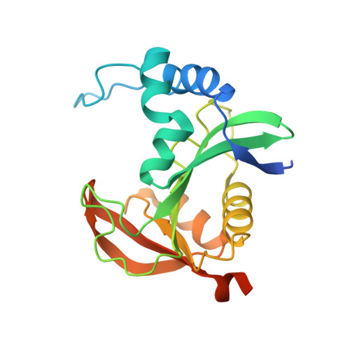
Enzyme structural plasticity and the emergence of broad-spectrum antibiotic resistance.

Maurice, F.Broutin, I.Podglajen, I.Benas, P.Collatz, E.Dardel, F.

(2008) EMBO Rep 9: 344-349

  • DOI: https://doi.org/10.1038/embor.2008.9
  • Primary Citation of Related Structures:  
    2PR8, 2PRB, 2QIR

  • PubMed Abstract: 

    The emergence of multi-resistant pathogenic bacteria is a worldwide health issue. Recently, clinical variants of a single antibiotic-modifying acetyltransferase, AAC(6')-Ib-a variant of aminoglycoside 6'-N-acetyltransferase-have been identified that confer extended resistance to most aminoglycosides and, more surprisingly, to structurally unrelated fluoroquinolones. The corresponding gene is carried by mobile genetic elements and is present in most multi-resistant pathogenic strains, hence making it a serious threat to current therapies. Here, we report the crystal structures of both narrow- and broad-spectrum resistance variants of this enzyme, which reveal the structural basis for the emergence of extended resistance. The active site shows an important plasticity and has adapted to new substrates by a large-scale gaping process. We have also obtained co-crystals with both substrates, and with a simple transition state analogue, which provides new clues for the design of inhibitors of this resistance mechanism.


  • Organizational Affiliation

    Cristallographie and RMN Biologiques, Université Paris Descartes, CNRS, 4 Avenue de l'Observatoire, 75006 Paris, France.


Macromolecules
Find similar proteins by:  (by identity cutoff)  |  3D Structure
Entity ID: 1
MoleculeChains Sequence LengthOrganismDetailsImage
Aminoglycoside 6-N-acetyltransferase type Ib11
A, B
196Salmonella enterica subsp. enterica serovar TyphimuriumMutation(s): 0 
EC: 2.3.1.82
UniProt
Find proteins for Q8GLI5 (Salmonella typhimurium)
Explore Q8GLI5 
Go to UniProtKB:  Q8GLI5
Entity Groups  
Sequence Clusters30% Identity50% Identity70% Identity90% Identity95% Identity100% Identity
UniProt GroupQ8GLI5
Sequence Annotations
Expand
  • Reference Sequence
Small Molecules
Ligands 1 Unique
IDChains Name / Formula / InChI Key2D Diagram3D Interactions
EPE
Query on EPE

Download Ideal Coordinates CCD File 
C [auth A],
D [auth B]
4-(2-HYDROXYETHYL)-1-PIPERAZINE ETHANESULFONIC ACID
C8 H18 N2 O4 S
JKMHFZQWWAIEOD-UHFFFAOYSA-N
Experimental Data & Validation

Experimental Data

  • Method: X-RAY DIFFRACTION
  • Resolution: 2.10 Å
  • R-Value Free: 0.252 
  • R-Value Work: 0.196 
  • R-Value Observed: 0.201 
  • Space Group: C 2 2 21
Unit Cell:
Length ( Å )Angle ( ˚ )
a = 71.62α = 90
b = 85.37β = 90
c = 150.41γ = 90
Software Package:
Software NamePurpose
XSCALEdata scaling
PHASERphasing
SHARPphasing
REFMACrefinement
PDB_EXTRACTdata extraction
ADSCdata collection
XDSdata reduction

Structure Validation

View Full Validation Report


Entry History 

Deposition Data

Revision History  (Full details and data files)

  • Version 1.0: 2008-04-08
    Type: Initial release
  • Version 1.1: 2011-07-13
    Changes: Version format compliance
  • Version 1.2: 2024-02-21
    Changes: Data collection, Database references, Derived calculations