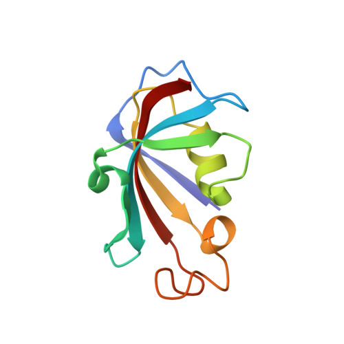
Structural coupling between FKBP12 and buried water.
Szep, S., Park, S., Boder, E.T., Van Duyne, G.D., Saven, J.G.(2009) Proteins 74: 603-611
- PubMed: 18704951
- DOI: https://doi.org/10.1002/prot.22176
- Primary Citation of Related Structures:
2PPN, 2PPO, 2PPP - PubMed Abstract:
Globular proteins often contain structurally well-resolved internal water molecules. Previously, we reported results from a molecular dynamics study that suggested that buried water (Wat3) may play a role in modulating the structure of the FK506 binding protein-12 (FKBP12) (Park and Saven, Proteins 2005; 60:450-463). In particular, simulations suggested that disrupting a hydrogen bond to Wat3 by mutating E60 to either A or Q would cause a structural perturbation involving the distant W59 side chain, which rotates to a new conformation in response to the mutation. This effectively remodels the ligand-binding pocket, as the side chain in the new conformation is likely to clash with bound FK506. To test whether the protein structure is in effect modulated by the binding of a buried water in the distance, we determined high-resolution (0.92-1.29 A) structures of wild-type FKBP12 and its two mutants (E60A, E60Q) by X-ray crystallography. The structures of mutant FKBP12 show that the ligand-binding pocket is indeed remodeled as predicted by the substitution at position 60, even though the water molecule does not directly interact with any of the amino acids of the binding pocket. Thus, these structures support the view that buried water molecules constitute an integral, noncovalent component of the protein structure. Additionally, this study provides an example in which predictions from molecular dynamics simulations are experimentally validated with atomic precision, thus showing that the structural features of protein-water interactions can be reliably modeled at a molecular level.
- Department of Biochemistry and Biophysics, Howard Hughes Medical Institute, University of Pennsylvania, Philadelphia, Pennsylvania 19104, USA.
Organizational Affiliation:


