2POR | pdb_00002por

STRUCTURE OF PORIN REFINED AT 1.8 ANGSTROMS RESOLUTION


Experimental Data Snapshot

  • Method: X-RAY DIFFRACTION
  • Resolution: 1.80 Å
  • R-Value Work: 
    0.186 (Depositor), 0.169 (DCC) 

wwPDB Validation 3D Report Full Report

Validation slider image for 2POR

Ligand Structure Quality Assessment 


This is version 1.5 of the entry. See complete history

Literature

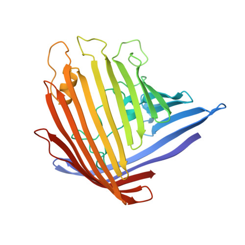
Structure of porin refined at 1.8 A resolution.

Weiss, M.S.Schulz, G.E.

(1992) J Mol Biology 227: 493-509

  • DOI: https://doi.org/10.1016/0022-2836(92)90903-w
  • Primary Citation Related Structures: 
    2POR

  • PubMed Abstract: 

    The crystal structure of porin from Rhodobacter capsulatus has been refined using the simulated annealing method. The final model consists of all 301 amino acid residues well obeying standard geometry, three calcium ions, 274 solvent molecules, three detergent molecules and one unknown ligand modeled as a detergent molecule. The final crystallographic R-factor is 18.6% based on 42,851 independent reflections in the resolution range 10 to 1.8 A. The model is described in detail.


  • Organizational Affiliation
    • Institut für Organische Chemie und Biochemie der Universität, Freiburg, Germany.

Macromolecule Content 

  • Total Structure Weight: 32.9 kDa 
  • Atom Count: 2,589 
  • Modeled Residue Count: 301 
  • Deposited Residue Count: 301 
  • Unique protein chains: 1

Macromolecules

Find similar proteins by:|  3D Structure
Entity ID: 1
MoleculeChains  Sequence LengthOrganismDetailsImage
PORIN301Rhodobacter capsulatusMutation(s): 0 
Membrane Entity: Yes 
UniProt
Find proteins for P31243 (Rhodobacter capsulatus)
Explore P31243 
Go to UniProtKB:  P31243
Entity Groups
Sequence Clusters30% Identity50% Identity70% Identity90% Identity95% Identity100% Identity
UniProt GroupP31243
Sequence Annotations
Expand
Reference Sequence

Experimental Data & Validation

Experimental Data

  • Method: X-RAY DIFFRACTION
  • Resolution: 1.80 Å
  • R-Value Work:  0.186 (Depositor), 0.169 (DCC) 
Space Group: H 3
Unit Cell:
Length ( Å )Angle ( ˚ )
a = 92.3α = 90
b = 92.3β = 90
c = 146.2γ = 120
Software Package:
Software NamePurpose
X-PLORrefinement

Structure Validation

View Full Validation Report


Ligand Structure Quality Assessment 


Entry History 

Deposition Data

Revision History  (Full details and data files)

  • Version 1.0: 1993-07-15
    Type: Initial release
  • Version 1.1: 2008-03-25
    Changes: Version format compliance
  • Version 1.2: 2011-07-13
    Changes: Derived calculations, Version format compliance
  • Version 1.3: 2011-11-16
    Changes: Atomic model
  • Version 1.4: 2017-11-29
    Changes: Derived calculations, Other
  • Version 1.5: 2024-02-21
    Changes: Data collection, Database references, Derived calculations