2POH

Structure of Phage P22 Tail Needle gp26


Experimental Data Snapshot

  • Method: X-RAY DIFFRACTION
  • Resolution: 2.10 Å
  • R-Value Free: 0.232 

wwPDB Validation   3D Report Full Report


This is version 1.2 of the entry. See complete history


Literature

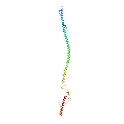
Structure of phage P22 cell envelope-penetrating needle.

Olia, A.S.Casjens, S.Cingolani, G.

(2007) Nat Struct Mol Biol 14: 1221-1226

  • DOI: https://doi.org/10.1038/nsmb1317
  • Primary Citation of Related Structures:  
    2POH

  • PubMed Abstract: 

    Bacteriophage P22 infects Salmonella enterica by injecting its genetic material through the cell envelope. During infection, a specialized tail needle, gp26, is injected into the host, likely piercing a hole in the host cell envelope. The 2.1-Å crystal structure of gp26 reveals a 240-Å elongated protein fiber formed by two trimeric coiled-coil domains interrupted by a triple β-helix.The N terminus of gp26 plugs the portal protein channel, retaining the genetic material inside the virion. The C-terminal tip of the fiber exposes β-hairpins with hydrophobic tips similar to those seen in class II fusion peptides. The α-helical core connecting these two functionally polarized tips presents four trimerization octads with consensus sequence IXXLXXXV. The slender conformation of the gp26 fiber minimizes the surface exposed to solvent, which is consistent with the idea that gp26 traverses the cell envelope lipid bilayers.


  • Organizational Affiliation

    Department of Biochemistry and Molecular Biology, SUNY Upstate Medical University, 750 East Adams Street, Syracuse, New York 13210, USA.


Macromolecules
Find similar proteins by:  (by identity cutoff)  |  3D Structure
Entity ID: 1
MoleculeChains Sequence LengthOrganismDetailsImage
Head completion protein
A, B, C, D, E
A, B, C, D, E, F
233Salmonella phage P22-pbiMutation(s): 2 
Gene Names: 26
UniProt
Find proteins for P35837 (Salmonella phage P22)
Explore P35837 
Go to UniProtKB:  P35837
Entity Groups  
Sequence Clusters30% Identity50% Identity70% Identity90% Identity95% Identity100% Identity
UniProt GroupP35837
Sequence Annotations
Expand
  • Reference Sequence
Small Molecules
Modified Residues  1 Unique
IDChains TypeFormula2D DiagramParent
MSE
Query on MSE
A, B, C, D, E
A, B, C, D, E, F
L-PEPTIDE LINKINGC5 H11 N O2 SeMET
Experimental Data & Validation

Experimental Data

  • Method: X-RAY DIFFRACTION
  • Resolution: 2.10 Å
  • R-Value Free: 0.232 
  • Space Group: P 1 21 1
Unit Cell:
Length ( Å )Angle ( ˚ )
a = 40.398α = 90
b = 114.029β = 90.74
c = 171.917γ = 90
Software Package:
Software NamePurpose
SHELXmodel building
SHELXL-97refinement
DENZOdata reduction
SCALEPACKdata scaling
SHELXphasing

Structure Validation

View Full Validation Report


Entry History 

Deposition Data

Revision History  (Full details and data files)

  • Version 1.0: 2007-12-04
    Type: Initial release
  • Version 1.1: 2011-07-13
    Changes: Version format compliance
  • Version 1.2: 2021-10-20
    Changes: Database references, Derived calculations