2PKQ

Crystal structure of the photosynthetic A2B2-glyceraldehyde-3-phosphate dehydrogenase, complexed with NADP


Experimental Data Snapshot

  • Method: X-RAY DIFFRACTION
  • Resolution: 3.60 Å
  • R-Value Free: 0.292 
  • R-Value Work: 0.261 
  • R-Value Observed: 0.261 

wwPDB Validation   3D Report Full Report


Ligand Structure Quality Assessment 


This is version 1.5 of the entry. See complete history


Literature

Molecular mechanism of thioredoxin regulation in photosynthetic A2B2-glyceraldehyde-3-phosphate dehydrogenase.

Fermani, S.Sparla, F.Falini, G.Martelli, P.L.Casadio, R.Pupillo, P.Ripamonti, A.Trost, P.

(2007) Proc Natl Acad Sci U S A 104: 11109-11114

  • DOI: https://doi.org/10.1073/pnas.0611636104
  • Primary Citation of Related Structures:  
    2PKQ, 2PKR

  • PubMed Abstract: 

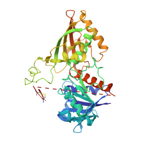
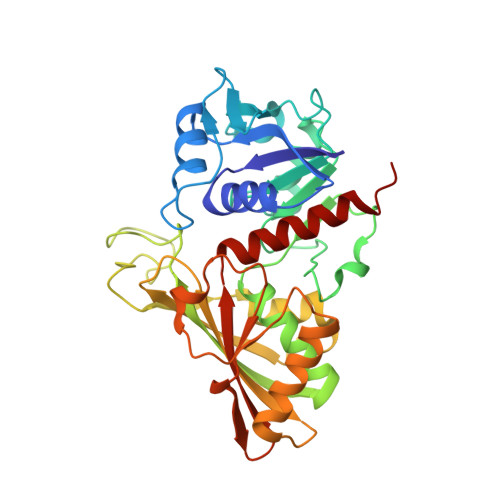
    Chloroplast glyceraldehyde-3-phosphate dehydrogenase (GAPDH) is a light-regulated, NAD(P)H-dependent enzyme involved in plant photosynthetic carbon reduction. Unlike lower photosynthetic organisms, which only contain A(4)-GAPDH, the major GAPDH isoform of land plants is made up of A and B subunits, the latter containing a C-terminal extension (CTE) with fundamental regulatory functions. Light-activation of AB-GAPDH depends on the redox state of a pair of cysteines of the CTE, which can form a disulfide bond under control of thioredoxin f, leading to specific inhibition of the NADPH-dependent activity. The tridimensional structure of A(2)B(2)-GAPDH from spinach chloroplasts, crystallized in the oxidized state, shows that each disulfide-containing CTE is docked into a deep cleft between a pair of A and B subunits. The structure of the CTE was derived from crystallographic data and computational modeling and confirmed by site-specific mutagenesis. Structural analysis of oxidized A(2)B(2)-GAPDH and chimeric mutant [A+CTE](4)-GAPDH revealed that Arg-77, which is essential for coenzyme specificity and high NADPH-dependent activity, fails to interact with NADP in these kinetically inhibited GAPDH tetramers and is attracted instead by negative residues of oxidized CTE. Other subtle changes in catalytic domains and overall conformation of the tetramers were noticed in oxidized A(2)B(2)-GAPDH and [A+CTE](4)-GAPDH, compared with fully active A(4)-GAPDH. The CTE is envisioned as a redox-sensitive regulatory domain that can force AB-GAPDH into a kinetically inhibited conformation under oxidizing conditions, which also occur during dark inactivation of the enzyme in vivo.


  • Organizational Affiliation

    Department of Chemistry, University of Bologna, Via Selmi 2, 40126 Bologna, Italy.


Macromolecules
Find similar proteins by:  (by identity cutoff)  |  3D Structure
Entity ID: 1
MoleculeChains Sequence LengthOrganismDetailsImage
Glyceraldehyde-3-phosphate dehydrogenase BA [auth O],
C [auth Q],
F [auth T]
368Spinacia oleraceaMutation(s): 0 
EC: 1.2.1.13
UniProt
Find proteins for P12860 (Spinacia oleracea)
Explore P12860 
Go to UniProtKB:  P12860
Entity Groups  
Sequence Clusters30% Identity50% Identity70% Identity90% Identity95% Identity100% Identity
UniProt GroupP12860
Sequence Annotations
Expand
  • Reference Sequence
Find similar proteins by:  (by identity cutoff)  |  3D Structure
Entity ID: 2
MoleculeChains Sequence LengthOrganismDetailsImage
Glyceraldehyde-3-phosphate dehydrogenase AB [auth P],
D [auth R],
E [auth S]
337Spinacia oleraceaMutation(s): 0 
EC: 1.2.1.13
UniProt
Find proteins for P19866 (Spinacia oleracea)
Explore P19866 
Go to UniProtKB:  P19866
Entity Groups  
Sequence Clusters30% Identity50% Identity70% Identity90% Identity95% Identity100% Identity
UniProt GroupP19866
Sequence Annotations
Expand
  • Reference Sequence
Small Molecules
Ligands 2 Unique
IDChains Name / Formula / InChI Key2D Diagram3D Interactions
NDP
Query on NDP

Download Ideal Coordinates CCD File 
AA [auth T]
H [auth O]
K [auth P]
O [auth Q]
S [auth R]
AA [auth T],
H [auth O],
K [auth P],
O [auth Q],
S [auth R],
W [auth S]
NADPH DIHYDRO-NICOTINAMIDE-ADENINE-DINUCLEOTIDE PHOSPHATE
C21 H30 N7 O17 P3
ACFIXJIJDZMPPO-NNYOXOHSSA-N
SO4
Query on SO4

Download Ideal Coordinates CCD File 
G [auth O]
I [auth P]
J [auth P]
L [auth Q]
M [auth Q]
G [auth O],
I [auth P],
J [auth P],
L [auth Q],
M [auth Q],
N [auth Q],
P [auth R],
Q [auth R],
R,
T [auth S],
U [auth S],
V [auth S],
X [auth T],
Y [auth T],
Z [auth T]
SULFATE ION
O4 S
QAOWNCQODCNURD-UHFFFAOYSA-L
Experimental Data & Validation

Experimental Data

  • Method: X-RAY DIFFRACTION
  • Resolution: 3.60 Å
  • R-Value Free: 0.292 
  • R-Value Work: 0.261 
  • R-Value Observed: 0.261 
  • Space Group: C 1 2 1
Unit Cell:
Length ( Å )Angle ( ˚ )
a = 186.147α = 90
b = 215.687β = 102.52
c = 81.469γ = 90
Software Package:
Software NamePurpose
CNSrefinement
MAR345data collection
DENZOdata reduction
SCALEPACKdata scaling
AMoREphasing

Structure Validation

View Full Validation Report


Ligand Structure Quality Assessment 


Entry History 

Deposition Data

Revision History  (Full details and data files)

  • Version 1.0: 2007-06-19
    Type: Initial release
  • Version 1.1: 2007-10-24
    Changes: Version format compliance
  • Version 1.2: 2011-07-13
    Changes: Derived calculations, Version format compliance
  • Version 1.3: 2014-11-12
    Changes: Structure summary
  • Version 1.4: 2017-10-18
    Changes: Refinement description
  • Version 1.5: 2023-08-30
    Changes: Data collection, Database references, Derived calculations, Refinement description