2PKK

Crystal structure of M tuberculosis Adenosine Kinase complexed with 2-fluro adenosine


Experimental Data Snapshot

  • Method: X-RAY DIFFRACTION
  • Resolution: 1.93 Å
  • R-Value Free: 0.240 
  • R-Value Work: 0.189 
  • R-Value Observed: 0.192 

wwPDB Validation   3D Report Full Report


Ligand Structure Quality Assessment 


This is version 1.3 of the entry. See complete history


Literature

High resolution crystal structures of Mycobacterium tuberculosis adenosine kinase: insights into the mechanism and specificity of this novel prokaryotic enzyme

Reddy, M.C.M.Palaninathan, S.K.Shetty, N.D.Owen, J.L.Watson, M.D.Sacchettini, J.C.

(2007) J Biol Chem 282: 27334-27342

  • DOI: https://doi.org/10.1074/jbc.M703290200
  • Primary Citation of Related Structures:  
    2PKF, 2PKK, 2PKM, 2PKN

  • PubMed Abstract: 

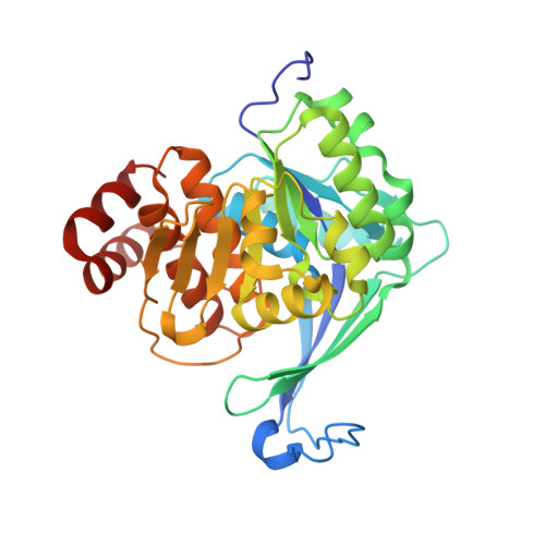
    Adenosine kinase (ADK) catalyzes the phosphorylation of adenosine (Ado) to adenosine monophosphate (AMP). It is part of the purine salvage pathway that has been identified only in eukaryotes, with the single exception of Mycobacterium spp. Whereas it is not clear if Mycobacterium tuberculosis (Mtb) ADK is essential, it has been shown that the enzyme can selectively phosphorylate nucleoside analogs to produce products toxic to the cell. We have determined the crystal structure of Mtb ADK unliganded as well as ligand (Ado) bound at 1.5- and 1.9-A resolution, respectively. The structure of the binary complexes with the inhibitor 2-fluoroadenosine (F-Ado) bound and with the adenosine 5'-(beta,gamma-methylene)triphosphate (AMP-PCP) (non-hydrolyzable ATP analog) bound were also solved at 1.9-A resolution. These four structures indicate that Mtb ADK is a dimer formed by an extended beta sheet. The active site of the unliganded ADK is in an open conformation, and upon Ado binding a lid domain of the protein undergoes a large conformation change to close the active site. In the closed conformation, the lid forms direct interactions with the substrate and residues of the active site. Interestingly, AMP-PCP binding alone was not sufficient to produce the closed state of the enzyme. The binding mode of F-Ado was characterized to illustrate the role of additional non-bonding interactions in Mtb ADK compared with human ADK.


  • Organizational Affiliation

    Department of Biochemistry & Biophysics, Texas A&M University, College Station, Texas 77843.


Macromolecules
Find similar proteins by:  (by identity cutoff)  |  3D Structure
Entity ID: 1
MoleculeChains Sequence LengthOrganismDetailsImage
Adenosine kinase334Mycobacterium tuberculosisMutation(s): 0 
Gene Names: adoK
EC: 2.7.1.20
UniProt
Find proteins for P9WID5 (Mycobacterium tuberculosis (strain ATCC 25618 / H37Rv))
Explore P9WID5 
Go to UniProtKB:  P9WID5
Entity Groups  
Sequence Clusters30% Identity50% Identity70% Identity90% Identity95% Identity100% Identity
UniProt GroupP9WID5
Sequence Annotations
Expand
  • Reference Sequence
Small Molecules
Ligands 1 Unique
IDChains Name / Formula / InChI Key2D Diagram3D Interactions
2FA
Query on 2FA

Download Ideal Coordinates CCD File 
B [auth A]2-(6-AMINO-2-FLUORO-PURIN-9-YL)-5-HYDROXYMETHYL-TETRAHYDRO-FURAN-3,4-DIOL
C10 H12 F N5 O4
HBUBKKRHXORPQB-UUOKFMHZSA-N
Experimental Data & Validation

Experimental Data

  • Method: X-RAY DIFFRACTION
  • Resolution: 1.93 Å
  • R-Value Free: 0.240 
  • R-Value Work: 0.189 
  • R-Value Observed: 0.192 
  • Space Group: P 31 2 1
Unit Cell:
Length ( Å )Angle ( ˚ )
a = 71.916α = 90
b = 71.916β = 90
c = 110.521γ = 120
Software Package:
Software NamePurpose
REFMACrefinement
CrystalCleardata collection
CrystalCleardata reduction
CrystalCleardata scaling
PHASERphasing

Structure Validation

View Full Validation Report


Ligand Structure Quality Assessment 


Entry History 

Revision History  (Full details and data files)

  • Version 1.0: 2007-06-19
    Type: Initial release
  • Version 1.1: 2008-01-31
    Changes: Version format compliance
  • Version 1.2: 2011-07-13
    Changes: Derived calculations, Version format compliance
  • Version 1.3: 2024-02-21
    Changes: Data collection, Database references, Derived calculations