2P86 | pdb_00002p86

The high resolution crystal structure of rhodesain, the major cathepsin L protease from T. brucei rhodesiense, bound to inhibitor K11002


Experimental Data Snapshot

  • Method: X-RAY DIFFRACTION
  • Resolution: 1.16 Å
  • R-Value Free: 
    0.130 (Depositor), 0.141 (DCC) 
  • R-Value Work: 
    0.110 (Depositor), 0.124 (DCC) 
  • R-Value Observed: 
    0.111 (Depositor) 

Starting Model: experimental
View more details

wwPDB Validation 3D Report Full Report

Validation slider image for 2P86

Ligand Structure Quality Assessment 


This is version 1.5 of the entry. See complete history

Literature

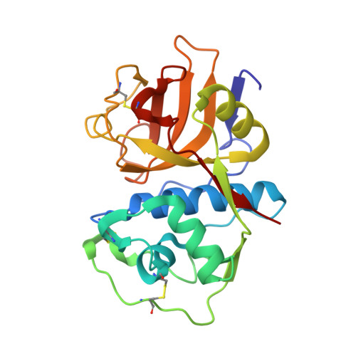
THE high resolution structure of rhodesain, the major cathepsin L protease from trypanosoma brucei rhodesiense, illustrates the basis for differences in inhibition profiles from other papain family cysteine proteases

Marion, R.Hansell, E.Caffrey, C.Roush, W.R.Brinen, L.S.

To be published.

Macromolecule Content 

  • Total Structure Weight: 24.15 kDa 
  • Atom Count: 2,135 
  • Modeled Residue Count: 215 
  • Deposited Residue Count: 215 
  • Unique protein chains: 1

Macromolecules

Find similar proteins by:|  3D Structure
Entity ID: 1
MoleculeChains  Sequence LengthOrganismDetailsImage
Cysteine protease215Trypanosoma brucei rhodesienseMutation(s): 1 
Gene Names: rhodesain
EC: 3.4.22.15
UniProt
Find proteins for Q95PM0 (Trypanosoma brucei rhodesiense)
Explore Q95PM0 
Go to UniProtKB:  Q95PM0
Entity Groups
Sequence Clusters30% Identity50% Identity70% Identity90% Identity95% Identity100% Identity
UniProt GroupQ95PM0
Sequence Annotations
Expand
Reference Sequence

Small Molecules

Ligands 2 Unique
IDChains Name / Formula / InChI Key2D Diagram3D Interactions
VS1

Query on VS1


Download:Ideal Coordinates CCD File
B [auth A]3-[[N-[MORPHOLIN-N-YL]-CARBONYL]-PHENYLALANINYL-AMINO]-5- PHENYL-PENTANE-1-SULFONYLBENZENE
C31 H37 N3 O5 S
NNOXYPYTWAQTTO-YTMVLYRLSA-N
EDO

Query on EDO


Download:Ideal Coordinates CCD File
C [auth A]
D [auth A]
E [auth A]
F [auth A]
G [auth A]
C [auth A],
D [auth A],
E [auth A],
F [auth A],
G [auth A],
H [auth A],
I [auth A],
J [auth A],
K [auth A],
L [auth A],
M [auth A]
1,2-ETHANEDIOL
C2 H6 O2
LYCAIKOWRPUZTN-UHFFFAOYSA-N

Experimental Data & Validation

Experimental Data

  • Method: X-RAY DIFFRACTION
  • Resolution: 1.16 Å
  • R-Value Free:  0.130 (Depositor), 0.141 (DCC) 
  • R-Value Work:  0.110 (Depositor), 0.124 (DCC) 
  • R-Value Observed: 0.111 (Depositor) 
Space Group: P 21 21 21
Unit Cell:
Length ( Å )Angle ( ˚ )
a = 33.659α = 90
b = 78.628β = 90
c = 80.725γ = 90
Software Package:
Software NamePurpose
REFMACrefinement
MOSFLMdata reduction
SCALAdata scaling
PHASERphasing

Structure Validation

View Full Validation Report


Ligand Structure Quality Assessment 


Entry History 

Deposition Data

Revision History  (Full details and data files)

  • Version 1.0: 2008-04-01
    Type: Initial release
  • Version 1.1: 2011-07-13
    Changes: Version format compliance
  • Version 1.2: 2018-10-24
    Changes: Data collection, Structure summary
  • Version 1.3: 2021-10-20
    Changes: Database references, Derived calculations
  • Version 1.4: 2023-08-30
    Changes: Data collection, Refinement description
  • Version 1.5: 2024-11-13
    Changes: Structure summary