2P81 | pdb_00002p81

Engrailed homeodomain helix-turn-helix motif


Experimental Data Snapshot

  • Method: SOLUTION NMR
  • Conformers Calculated: 50 
  • Conformers Submitted: 25 
  • Selection Criteria: structures with the lowest energy 

wwPDB Validation 3D Report Full Report

Validation slider image for 2P81

This is version 1.4 of the entry. See complete history

Literature

The helix-turn-helix motif as an ultrafast independently folding domain: The pathway of folding of Engrailed homeodomain.

Religa, T.L.Johnson, C.M.Vu, D.M.Brewer, S.H.Dyer, R.B.Fersht, A.R.

(2007) Proc Natl Acad Sci U S A 104: 9272-9277

  • DOI: https://doi.org/10.1073/pnas.0703434104
  • Primary Citation Related Structures: 
    2P81

  • PubMed Abstract: 

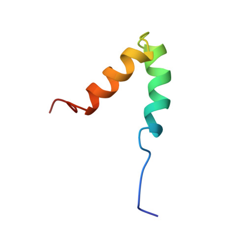
    Helices 2 and 3 of Engrailed homeodomain (EnHD) form a helix-turn-helix (HTH) motif. This common motif is believed not to fold independently, which is the characteristic feature of a motif rather than a domain. But we found that the EnHD HTH motif is monomeric and folded in solution, having essentially the same structure as in full-length protein. It had a sigmoidal thermal denaturation transition. Both native backbone and local tertiary interactions were formed concurrently at 4 x 10(5) s(-1) at 25 degrees C, monitored by IR and fluorescence T-jump kinetics, respectively, the same rate constant as for the fast phase in the folding of EnHD. The HTH motif, thus, is an ultrafast-folding, natural protein domain. Its independent stability and appropriate folding kinetics account for the stepwise folding of EnHD, satisfy fully the criteria for an on-pathway intermediate, and explain the changes in mechanism of folding across the homeodomain family. Experiments on mutated and engineered fragments of the parent protein with different probes allowed the assignment of the observed kinetic phases to specific events to show that EnHD is not an example of one-state downhill folding.


  • Organizational Affiliation
    • Medical Research Council Centre for Protein Engineering, Hills Road, Cambridge, United Kingdom.

Macromolecule Content 

  • Total Structure Weight: 5.42 kDa 
  • Atom Count: 382 
  • Modeled Residue Count: 44 
  • Deposited Residue Count: 44 
  • Unique protein chains: 1

Macromolecules

Find similar proteins by:|  3D Structure
Entity ID: 1
MoleculeChains  Sequence LengthOrganismDetailsImage
Segmentation polarity homeobox protein engrailed44Drosophila melanogasterMutation(s): 1 
Gene Names: en
UniProt
Find proteins for P02836 (Drosophila melanogaster)
Explore P02836 
Go to UniProtKB:  P02836
Entity Groups
Sequence Clusters30% Identity50% Identity70% Identity90% Identity95% Identity100% Identity
UniProt GroupP02836
Sequence Annotations
Expand
Reference Sequence

Experimental Data & Validation

Experimental Data

  • Method: SOLUTION NMR
  • Conformers Calculated: 50 
  • Conformers Submitted: 25 
  • Selection Criteria: structures with the lowest energy 

Structure Validation

View Full Validation Report


Entry History 

Deposition Data

  • Released Date: 2007-06-12 
  • Deposition Author(s): Religa, T.L.

Revision History  (Full details and data files)

  • Version 1.0: 2007-06-12
    Type: Initial release
  • Version 1.1: 2008-05-01
    Changes: Version format compliance
  • Version 1.2: 2011-07-13
    Changes: Version format compliance
  • Version 1.3: 2021-10-20
    Changes: Database references, Derived calculations
  • Version 1.4: 2024-05-22
    Changes: Data collection