2P5M

C-terminal domain hexamer of AhrC bound with L-arginine


Experimental Data Snapshot

  • Method: X-RAY DIFFRACTION
  • Resolution: 1.95 Å
  • R-Value Free: 0.224 
  • R-Value Work: 0.182 
  • R-Value Observed: 0.187 

wwPDB Validation   3D Report Full Report


Ligand Structure Quality Assessment 


This is version 1.3 of the entry. See complete history


Literature

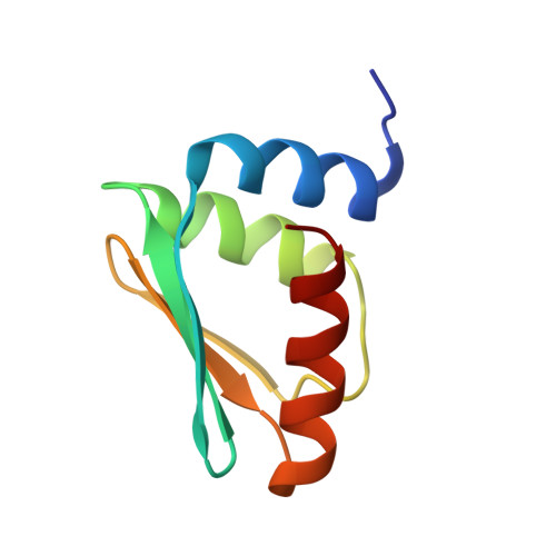
Structure of the C-terminal effector-binding domain of AhrC bound to its corepressor L-arginine.

Garnett, J.A.Baumberg, S.Stockley, P.G.Phillips, S.E.

(2007) Acta Crystallogr Sect F Struct Biol Cryst Commun 63: 918-921

  • DOI: https://doi.org/10.1107/S1744309107049391
  • Primary Citation of Related Structures:  
    2P5M

  • PubMed Abstract: 

    The arginine repressor/activator protein (AhrC) from Bacillus subtilis belongs to a large family of multifunctional transcription factors that are involved in the regulation of bacterial arginine metabolism. AhrC interacts with operator sites in the promoters of arginine biosynthetic and catabolic operons, acting as a transcriptional repressor at biosynthetic sites and an activator of transcription at catabolic sites. AhrC is a hexamer of identical subunits, each having two domains. The C-terminal domains form the core of the protein and are involved in oligomerization and L-arginine binding. The N-terminal domains lie on the outside of the compact core and play a role in binding to 18 bp DNA operators called ARG boxes. The C-terminal domain of AhrC has been expressed, purified and characterized, and also crystallized as a hexamer with the bound corepressor L-arginine. Here, the crystal structure refined to 1.95 A is presented.


  • Organizational Affiliation

    Astbury Centre for Structural Molecular Biology, University of Leeds, Leeds LS2 9JT, England.


Macromolecules
Find similar proteins by:  (by identity cutoff)  |  3D Structure
Entity ID: 1
MoleculeChains Sequence LengthOrganismDetailsImage
Arginine repressor
A, B, C
83Bacillus subtilisMutation(s): 1 
Gene Names: argRahrC
UniProt
Find proteins for P17893 (Bacillus subtilis (strain 168))
Explore P17893 
Go to UniProtKB:  P17893
Entity Groups  
Sequence Clusters30% Identity50% Identity70% Identity90% Identity95% Identity100% Identity
UniProt GroupP17893
Sequence Annotations
Expand
  • Reference Sequence
Experimental Data & Validation

Experimental Data

  • Method: X-RAY DIFFRACTION
  • Resolution: 1.95 Å
  • R-Value Free: 0.224 
  • R-Value Work: 0.182 
  • R-Value Observed: 0.187 
  • Space Group: P 65 2 2
Unit Cell:
Length ( Å )Angle ( ˚ )
a = 117.014α = 90
b = 117.014β = 90
c = 68.927γ = 120
Software Package:
Software NamePurpose
REFMACrefinement
PDB_EXTRACTdata extraction
ADSCdata collection
MOSFLMdata reduction
SCALAdata scaling
AMoREphasing

Structure Validation

View Full Validation Report


Ligand Structure Quality Assessment 


Entry History 

Deposition Data

Revision History  (Full details and data files)

  • Version 1.0: 2007-10-30
    Type: Initial release
  • Version 1.1: 2011-07-13
    Changes: Version format compliance
  • Version 1.2: 2021-10-20
    Changes: Database references, Derived calculations
  • Version 1.3: 2023-08-30
    Changes: Data collection, Refinement description