Characterization of Salmonella typhimurium YegS, a putative lipid kinase homologous to eukaryotic sphingosine and diacylglycerol kinases
Nichols, C.E., Lamb, H.K., Lockyer, M., Charles, I.G., Pyne, S., Hawkins, A.R., Stammers, D.K.(2007) Proteins 68: 13-25
- PubMed: 17393457 Search on PubMed
- DOI: https://doi.org/10.1002/prot.21386
- Primary Citation Related Structures:
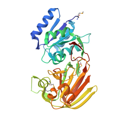
2P1R - PubMed Abstract:
Salmonella typhimurium YegS is a protein conserved in many prokaryotes. Although the function of YegS is not definitively known, it has been annotated as a potential diacylglycerol or sphingosine kinase based on sequence similarity with eukaryotic enzymes of known function. To further characterize YegS, we report its purification, biochemical analysis, crystallization, and structure determination. The crystal structure of YegS reveals a two-domain fold related to bacterial polyphosphate/ATP NAD kinases, comprising a central cleft between an N-terminal alpha/beta domain and a C-terminal two-layer beta-sandwich domain; conserved structural features are consistent with nucleotide binding within the cleft. The N-terminal and C-terminal domains of YegS are however counter-rotated, relative to the polyphosphate/ATP NAD kinase archetype, such that the potential nucleotide binding site is blocked. There are also two Ca2+ binding sites and two hydrophobic clefts, one in each domain of YegS. Analysis of mutagenesis data from eukaryotic homologues of YegS suggest that the N-terminal cleft may bind activating lipids while the C-terminal cleft may bind the lipid substrate. Microcalorimetry experiments showed interaction between recombinant YegS and Mg2+, Ca2+, and Mn2+ ions, with a weaker interaction also observed with polyphosphates and ATP. However, biochemical assays showed that recombinant YegS is endogenously neither an active diacylglycerol nor sphingosine kinase. Thus although the bioinformatics analysis and structure of YegS indicate that many of the ligand recognition determinants for lipid kinase activity are present, the absence of such activity may be due to specificity for a different lipid substrate or the requirement for activation by an, as yet, undetermined mechanism. In this regard the specific interaction of YegS with the periplasmic chaperone OmpH, which we demonstrate from pulldown experiments, may be of significance. Such an interaction suggests that YegS can be translocated to the periplasm and directed to the outer-membrane, an environment that may be required for enzyme activity.
- Division of Structural Biology, The Wellcome Trust Centre for Human Genetics, University of Oxford,Oxford OX3 7BN, United Kingdom.
Organizational Affiliation: