


Structures of actinonin bound peptide deformylases from E. faecalis and S. pyogenes
Kim, E.E., Kim, K.-H., Moon, J.H., Choi, K., Lee, H.K., Park, H.S.To be published.
Experimental Data Snapshot
Starting Model: experimental
View more details
Entity ID: 1 | |||||
|---|---|---|---|---|---|
| Molecule | Chains | Sequence Length | Organism | Details | Image |
| Peptide deformylase | 205 | Streptococcus pyogenes M1 GAS | Mutation(s): 0 Gene Names: def EC: 3.5.1.88 | 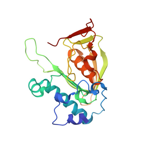 | |
UniProt | |||||
Entity Groups | |||||
| Sequence Clusters | 30% Identity50% Identity70% Identity90% Identity95% Identity100% Identity | ||||
| UniProt Group | P68771 | ||||
Sequence AnnotationsExpand | |||||
Reference Sequence | |||||
| Ligands 2 Unique | |||||
|---|---|---|---|---|---|
| ID | Chains | Name / Formula / InChI Key | 2D Diagram | 3D Interactions | |
| BB2 Download:Ideal Coordinates CCD File | C [auth A] | ACTINONIN C19 H35 N3 O5 XJLATMLVMSFZBN-VYDXJSESSA-N |  | ||
| CO Download:Ideal Coordinates CCD File | B [auth A] | COBALT (II) ION Co XLJKHNWPARRRJB-UHFFFAOYSA-N |  | ||
| Length ( Å ) | Angle ( ˚ ) |
|---|---|
| a = 40.557 | α = 90 |
| b = 40.557 | β = 90 |
| c = 217.938 | γ = 120 |
| Software Name | Purpose |
|---|---|
| CNS | refinement |
| HKL-2000 | data collection |
| HKL-2000 | data reduction |
| SCALEPACK | data scaling |
| CNS | phasing |


