


Crystal Structure of Penicillin V acylase from Bacillus subtilis
Suresh, C.G., Rathinaswamy, P., Pundle, A.V., Prabhune, A.A., Sivaraman, H., Brannigan, J.A., Dodson, G.G.To be published.
Experimental Data Snapshot
Starting Model: experimental
View more details
wwPDB Validation 3D Report Full Report

Entity ID: 1 | |||||
|---|---|---|---|---|---|
| Molecule | Chains | Sequence Length | Organism | Details | Image |
| Penicillin V acylase | 327 | Bacillus subtilis | Mutation(s): 0 Gene Names: yxeI EC: 3.5.1.11 | 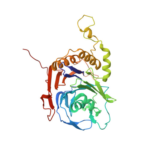 | |
UniProt | |||||
Entity Groups | |||||
| Sequence Clusters | 30% Identity50% Identity70% Identity90% Identity95% Identity100% Identity | ||||
| UniProt Group | P54948 | ||||
Sequence AnnotationsExpand | |||||
Reference Sequence | |||||
| Length ( Å ) | Angle ( ˚ ) |
|---|---|
| a = 110.963 | α = 90 |
| b = 307.956 | β = 90 |
| c = 56.003 | γ = 90 |
| Software Name | Purpose |
|---|---|
| REFMAC | refinement |
| CrystalClear | data collection |
| DENZO | data reduction |
| SCALEPACK | data scaling |
| AMoRE | phasing |


