2OLV | pdb_00002olv

Structural Insight Into the Transglycosylation Step Of Bacterial Cell Wall Biosynthesis : Donor Ligand Complex


Experimental Data Snapshot

  • Method: X-RAY DIFFRACTION
  • Resolution: 2.80 Å
  • R-Value Free: 
    0.280 (Depositor), 0.283 (DCC) 
  • R-Value Work: 
    0.231 (Depositor), 0.237 (DCC) 
  • R-Value Observed: 
    0.234 (Depositor) 

wwPDB Validation 3D Report Full Report

Validation slider image for 2OLV

Ligand Structure Quality Assessment 


This is version 1.4 of the entry. See complete history

Literature

Structural insight into the transglycosylation step of bacterial cell-wall biosynthesis.

Lovering, A.L.de Castro, L.H.Lim, D.Strynadka, N.C.

(2007) Science 315: 1402-1405

  • DOI: https://doi.org/10.1126/science.1136611
  • Primary Citation Related Structures: 
    2OLU, 2OLV

  • PubMed Abstract: 

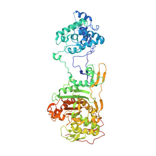
    Peptidoglycan glycosyltransferases (GTs) catalyze the polymerization step of cell-wall biosynthesis, are membrane-bound, and are highly conserved across all bacteria. Long considered the "holy grail" of antibiotic research, they represent an essential and easily accessible drug target for antibiotic-resistant bacteria, including methicillin-resistant Staphylococcus aureus. We have determined the 2.8 angstrom structure of a bifunctional cell-wall cross-linking enzyme, including its transpeptidase and GT domains, both unliganded and complexed with the substrate analog moenomycin. The peptidoglycan GTs adopt a fold distinct from those of other GT classes. The structures give insight into critical features of the catalytic mechanism and key interactions required for enzyme inhibition.


  • Organizational Affiliation
    • Department of Biochemistry and Molecular Biology, and Center for Blood Research, University of British Columbia, 2350 Health Sciences Mall, Vancouver, Canada.

Macromolecule Content 

  • Total Structure Weight: 152.44 kDa 
  • Atom Count: 9,878 
  • Modeled Residue Count: 1,235 
  • Deposited Residue Count: 1,338 
  • Unique protein chains: 1

Macromolecules

Find similar proteins by:|  3D Structure
Entity ID: 1
MoleculeChains  Sequence LengthOrganismDetailsImage
Penicillin-binding protein 2
A, B
669Staphylococcus aureusMutation(s): 14 
Gene Names: pbp2
EC: 2.3.2
Membrane Entity: Yes 
UniProt
Find proteins for Q9R744 (Staphylococcus aureus)
Explore Q9R744 
Go to UniProtKB:  Q9R744
Entity Groups
Sequence Clusters30% Identity50% Identity70% Identity90% Identity95% Identity100% Identity
UniProt GroupQ9R744
Sequence Annotations
Expand
Reference Sequence

Small Molecules

Modified Residues  1 Unique
IDChains TypeFormula2D DiagramParent
MSE
Query on MSE
A, B
L-PEPTIDE LINKINGC5 H11 N O2 SeMET

Experimental Data & Validation

Experimental Data

  • Method: X-RAY DIFFRACTION
  • Resolution: 2.80 Å
  • R-Value Free:  0.280 (Depositor), 0.283 (DCC) 
  • R-Value Work:  0.231 (Depositor), 0.237 (DCC) 
  • R-Value Observed: 0.234 (Depositor) 
Space Group: P 21 21 2
Unit Cell:
Length ( Å )Angle ( ˚ )
a = 79.565α = 90
b = 212.211β = 90
c = 91.635γ = 90
Software Package:
Software NamePurpose
SCALAdata scaling
REFMACrefinement
PDB_EXTRACTdata extraction
ADSCdata collection
MOSFLMdata reduction
CCP4data scaling
SOLVEphasing

Structure Validation

View Full Validation Report


Ligand Structure Quality Assessment 


Entry History 

Deposition Data

Revision History  (Full details and data files)

  • Version 1.0: 2007-03-13
    Type: Initial release
  • Version 1.1: 2008-05-01
    Changes: Version format compliance
  • Version 1.2: 2011-07-13
    Changes: Advisory, Version format compliance
  • Version 1.3: 2023-12-27
    Changes: Data collection, Database references, Derived calculations, Refinement description
  • Version 1.4: 2024-11-13
    Changes: Structure summary