2OK7

Ferredoxin-NADP+ reductase from Plasmodium falciparum with 2'P-AMP


Experimental Data Snapshot

  • Method: X-RAY DIFFRACTION
  • Resolution: 2.70 Å
  • R-Value Free: 0.321 
  • R-Value Work: 0.260 
  • R-Value Observed: 0.263 

wwPDB Validation   3D Report Full Report


Ligand Structure Quality Assessment 


This is version 1.3 of the entry. See complete history


Literature

Ferredoxin-NADP(+) Reductase from Plasmodium falciparum Undergoes NADP(+)-dependent Dimerization and Inactivation: Functional and Crystallographic Analysis.

Milani, M.Balconi, E.Aliverti, A.Mastrangelo, E.Seeber, F.Bolognesi, M.Zanetti, G.

(2007) J Mol Biol 367: 501-513

  • DOI: https://doi.org/10.1016/j.jmb.2007.01.005
  • Primary Citation of Related Structures:  
    2OK7, 2OK8

  • PubMed Abstract: 

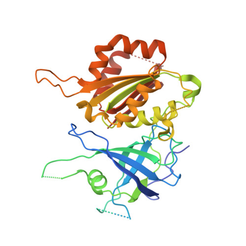
    The completion of the Plasmodium falciparum genome sequence has recently promoted the search for new antimalarial drugs. More specifically, metabolic pathways of the apicoplast, a key organelle for survival of the parasite, have been recognized as potential targets for the development of specific new antimalarial agents. As most apicomplexan parasites, P. falciparum displays a plant-type ferredoxin-NADP(+) reductase, yielding reduced ferredoxin for essential biosynthetic pathways in the apicoplast. Here we report a molecular, kinetic and ligand binding characterization of the recombinant ferredoxin-NADP(+) reductase from P. falciparum, in the light of current data available for plant ferredoxin-NADP(+) reductases. In parallel with the functional characterization, we describe the crystal structures of P. falciparum ferredoxin-NADP(+) reductase in free form and in complex with 2'-phospho-AMP (at 2.4 and 2.7 A resolution, respectively). The enzyme displays structural properties likely to be unique to plasmodial reductases. In particular, the two crystal structures highlight a covalent dimer, which relies on the oxidation of residue Cys99 in two opposing subunits, and a helix-coil transition that occurs in the NADP-binding domain, triggered by 2'-phospho-AMP binding. Studies in solution show that NADP(+), as well as 2'-phospho-AMP, promotes the formation of the disulfide-stabilized dimer. The isolated dimer is essentially inactive, but full activity is recovered upon disulfide reduction. The occurrence of residues unique to the plasmodial enzyme, and the discovery of specific conformational properties, highlight the NADP-binding domain of P. falciparum ferredoxin-NADP(+) reductase as particularly suited for the rational development of antimalarial compounds.


  • Organizational Affiliation

    CNR-INFM, Department of Biomolecular Sciences and Biotechnology, University of Milano, Via Celoria 26, 20133-Milano, Italy.


Macromolecules
Find similar proteins by:  (by identity cutoff)  |  3D Structure
Entity ID: 1
MoleculeChains Sequence LengthOrganismDetailsImage
Putative ferredoxin--NADP reductase
A, B, C, D, E
A, B, C, D, E, F
316Plasmodium falciparum 3D7Mutation(s): 0 
Gene Names: ORF PFF1115w
EC: 1.18.1.2
UniProt
Find proteins for C6KT68 (Plasmodium falciparum (isolate 3D7))
Explore C6KT68 
Go to UniProtKB:  C6KT68
Entity Groups  
Sequence Clusters30% Identity50% Identity70% Identity90% Identity95% Identity100% Identity
UniProt GroupC6KT68
Sequence Annotations
Expand
  • Reference Sequence
Small Molecules
Ligands 3 Unique
IDChains Name / Formula / InChI Key2D Diagram3D Interactions
FAD
Query on FAD

Download Ideal Coordinates CCD File 
H [auth A]
J [auth B]
L [auth C]
N [auth D]
P [auth E]
H [auth A],
J [auth B],
L [auth C],
N [auth D],
P [auth E],
R [auth F]
FLAVIN-ADENINE DINUCLEOTIDE
C27 H33 N9 O15 P2
VWWQXMAJTJZDQX-UYBVJOGSSA-N
A2P
Query on A2P

Download Ideal Coordinates CCD File 
I [auth A]
K [auth B]
M [auth C]
O [auth D]
Q [auth E]
I [auth A],
K [auth B],
M [auth C],
O [auth D],
Q [auth E],
S [auth F]
ADENOSINE-2'-5'-DIPHOSPHATE
C10 H15 N5 O10 P2
AEOBEOJCBAYXBA-KQYNXXCUSA-N
NA
Query on NA

Download Ideal Coordinates CCD File 
G [auth A]SODIUM ION
Na
FKNQFGJONOIPTF-UHFFFAOYSA-N
Experimental Data & Validation

Experimental Data

  • Method: X-RAY DIFFRACTION
  • Resolution: 2.70 Å
  • R-Value Free: 0.321 
  • R-Value Work: 0.260 
  • R-Value Observed: 0.263 
  • Space Group: P 31
Unit Cell:
Length ( Å )Angle ( ˚ )
a = 123.129α = 90
b = 123.129β = 90
c = 133.807γ = 120
Software Package:
Software NamePurpose
MOLREPphasing
REFMACrefinement
MOSFLMdata reduction
CCP4data scaling

Structure Validation

View Full Validation Report


Ligand Structure Quality Assessment 


Entry History 

Deposition Data

Revision History  (Full details and data files)

  • Version 1.0: 2007-02-13
    Type: Initial release
  • Version 1.1: 2008-05-01
    Changes: Version format compliance
  • Version 1.2: 2011-07-13
    Changes: Version format compliance
  • Version 1.3: 2023-08-30
    Changes: Data collection, Database references, Derived calculations, Refinement description