2OGW | pdb_00002ogw

Structure of ABC type zinc transporter from E. coli


Experimental Data Snapshot

  • Method: X-RAY DIFFRACTION
  • Resolution: 1.83 Å
  • R-Value Free: 
    0.269 (Depositor), 0.264 (DCC) 
  • R-Value Work: 
    0.223 (Depositor), 0.220 (DCC) 
  • R-Value Observed: 
    0.225 (Depositor) 

wwPDB Validation 3D Report Full Report

Validation slider image for 2OGW

This is version 1.4 of the entry. See complete history

Literature

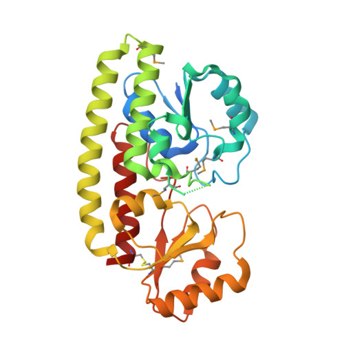
Structural Analysis of ABC-family Periplasmic Zinc Binding Protein Provides New Insights Into Mechanism of Ligand Uptake and Release

Chandra, B.R.Yogavel, M.Sharma, A.

(2007) J Mol Biology 367: 970-982

  • DOI: https://doi.org/10.1016/j.jmb.2007.01.041
  • Primary Citation Related Structures: 
    2OGW

  • PubMed Abstract: 

    ATP-binding cassette superfamily of periplasmic metal transporters are known to be vital for maintaining ion homeostasis in several pathogenic and non-pathogenic bacteria. We have determined crystal structure of the high-affinity zinc transporter ZnuA from Escherichia coli at 1.8 A resolution. This structure represents the first native (non-recombinant) protein structure of a periplasmic metal binding protein. ZnuA reveals numerous conformational features, which occur either in Zn(2+) or in Mn(2+) transporters, and presents a unique conformational state. A comprehensive comparison of ZnuA with other periplasmic ligand binding protein structures suggests vital mechanistic differences between bound and release states of metal transporters. The key new attributes in ZnuA include a C-domain disulfide bond, an extra alpha-helix proximal to the highly charged metal chelating mobile loop region, alternate conformations of secondary shell stabilizing residues at the metal binding site, and domain movements potentially controlled by salt bridges. Based on in-depth structural analyses of five metal binding transporters, we present here a mechanistic model termed as "partial domain slippage" for binding and release of Zn(2+).


  • Organizational Affiliation
    • Structural and Computational Biology Group, International Centre for Genetic Engineering and Biotechnology, New Delhi, India.

Macromolecule Content 

  • Total Structure Weight: 68.43 kDa 
  • Atom Count: 4,698 
  • Modeled Residue Count: 520 
  • Deposited Residue Count: 620 
  • Unique protein chains: 1

Macromolecules

Find similar proteins by:|  3D Structure
Entity ID: 1
MoleculeChains  Sequence LengthOrganismDetailsImage
High-affinity zinc uptake system protein znuA precursor
A, B
310Escherichia coliMutation(s): 7 
UniProt
Find proteins for P39172 (Escherichia coli (strain K12))
Explore P39172 
Go to UniProtKB:  P39172
Entity Groups
Sequence Clusters30% Identity50% Identity70% Identity90% Identity95% Identity100% Identity
UniProt GroupP39172
Sequence Annotations
Expand
Reference Sequence

Small Molecules

Modified Residues  1 Unique
IDChains TypeFormula2D DiagramParent
MSE
Query on MSE
A, B
L-PEPTIDE LINKINGC5 H11 N O2 SeMET

Experimental Data & Validation

Experimental Data

  • Method: X-RAY DIFFRACTION
  • Resolution: 1.83 Å
  • R-Value Free:  0.269 (Depositor), 0.264 (DCC) 
  • R-Value Work:  0.223 (Depositor), 0.220 (DCC) 
  • R-Value Observed: 0.225 (Depositor) 
Space Group: P 21 21 21
Unit Cell:
Length ( Å )Angle ( ˚ )
a = 72.9α = 90
b = 86.378β = 90
c = 87.99γ = 90
Software Package:
Software NamePurpose
REFMACrefinement
HKL-2000data collection
HKL-2000data reduction
HKL-2000data scaling
SOLVEphasing

Structure Validation

View Full Validation Report


Entry History 

Deposition Data

Revision History  (Full details and data files)

  • Version 1.0: 2007-03-06
    Type: Initial release
  • Version 1.1: 2008-05-01
    Changes: Version format compliance
  • Version 1.2: 2011-07-13
    Changes: Version format compliance
  • Version 1.3: 2023-12-27
    Changes: Data collection, Database references, Derived calculations
  • Version 1.4: 2024-10-30
    Changes: Structure summary