2OAA

Restriction endonuclease MvaI-cognate DNA substrate complex


Experimental Data Snapshot

  • Method: X-RAY DIFFRACTION
  • Resolution: 1.50 Å
  • R-Value Free: 0.194 
  • R-Value Work: 0.176 
  • R-Value Observed: 0.177 

wwPDB Validation   3D Report Full Report


This is version 1.4 of the entry. See complete history


Literature

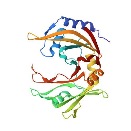
Restriction endonuclease MvaI is a monomer that recognizes its target sequence asymmetrically.

Kaus-Drobek, M.Czapinska, H.Sokolowska, M.Tamulaitis, G.Szczepanowski, R.H.Urbanke, C.Siksnys, V.Bochtler, M.

(2007) Nucleic Acids Res 35: 2035-2046

  • DOI: https://doi.org/10.1093/nar/gkm064
  • Primary Citation of Related Structures:  
    2OA9, 2OAA

  • PubMed Abstract: 

    Restriction endonuclease MvaI recognizes the sequence CC/WGG (W stands for A or T, '/' designates the cleavage site) and generates products with single nucleotide 5'-overhangs. The enzyme has been noted for its tolerance towards DNA modifications. Here, we report a biochemical characterization and crystal structures of MvaI in an apo-form and in a complex with target DNA at 1.5 A resolution. Our results show that MvaI is a monomer and recognizes its pseudosymmetric target sequence asymmetrically. The enzyme consists of two lobes. The catalytic lobe anchors the active site residues Glu36, Asp50, Glu55 and Lys57 and contacts the bases from the minor grove side. The recognition lobe mediates all major grove interactions with the bases. The enzyme in the crystal is bound to the strand with T at the center of the recognition sequence. The crystal structure with calcium ions and DNA mimics the prereactive state. MvaI shows structural similarities to BcnI, which cleaves the related sequence CC/SGG and to MutH enzyme, which is a component of the DNA repair machinery, and nicks one DNA strand instead of making a double-strand break.


  • Organizational Affiliation

    International Institute of Molecular and Cell Biology, Warsaw, Poland.


Macromolecules

Find similar proteins by:  (by identity cutoff)  |  3D Structure
Entity ID: 3
MoleculeChains Sequence LengthOrganismDetailsImage
R.MvaIE [auth A],
F [auth B]
249Kocuria variansMutation(s): 0 
Gene Names: mvaIR
UniProt
Find proteins for Q8RNV5 (Kocuria varians)
Explore Q8RNV5 
Go to UniProtKB:  Q8RNV5
Entity Groups  
Sequence Clusters30% Identity50% Identity70% Identity90% Identity95% Identity100% Identity
UniProt GroupQ8RNV5
Sequence Annotations
Expand
  • Reference Sequence

Find similar nucleic acids by:  Sequence   |   3D Structure  

Entity ID: 1
MoleculeChains LengthOrganismImage
5'-D(*GP*GP*TP*AP*CP*CP*TP*GP*GP*AP*T)-3'A [auth C],
C [auth E]
11N/A
Sequence Annotations
Expand
  • Reference Sequence

Find similar nucleic acids by:  Sequence   |   3D Structure  

Entity ID: 2
MoleculeChains LengthOrganismImage
5'-D(*CP*AP*TP*CP*CP*AP*GP*GP*TP*AP*C)-3'B [auth D],
D [auth F]
11N/A
Sequence Annotations
Expand
  • Reference Sequence
Experimental Data & Validation

Experimental Data

  • Method: X-RAY DIFFRACTION
  • Resolution: 1.50 Å
  • R-Value Free: 0.194 
  • R-Value Work: 0.176 
  • R-Value Observed: 0.177 
  • Space Group: P 1 21 1
Unit Cell:
Length ( Å )Angle ( ˚ )
a = 53.848α = 90
b = 81.38β = 90.52
c = 71.308γ = 90
Software Package:
Software NamePurpose
REFMACrefinement
MAR345data collection
MOSFLMdata reduction
CCP4data scaling
MOLREPphasing
NCSREFphasing

Structure Validation

View Full Validation Report


Entry History 

Deposition Data

Revision History  (Full details and data files)

  • Version 1.0: 2007-02-20
    Type: Initial release
  • Version 1.1: 2008-05-01
    Changes: Version format compliance
  • Version 1.2: 2011-07-13
    Changes: Advisory, Version format compliance
  • Version 1.3: 2017-10-18
    Changes: Advisory, Refinement description
  • Version 1.4: 2023-08-30
    Changes: Advisory, Data collection, Database references, Derived calculations, Refinement description