2OA8 | pdb_00002oa8

Crystal Structure of mTREX1 with ssDNA


Experimental Data Snapshot

  • Method: X-RAY DIFFRACTION
  • Resolution: 2.10 Å
  • R-Value Free: 
    0.259 (Depositor), 0.260 (DCC) 
  • R-Value Work: 
    0.192 (Depositor), 0.203 (DCC) 
  • R-Value Observed: 
    0.195 (Depositor) 

Starting Model: experimental
View more details

wwPDB Validation 3D Report Full Report

Validation slider image for 2OA8

This is version 1.5 of the entry. See complete history

Literature

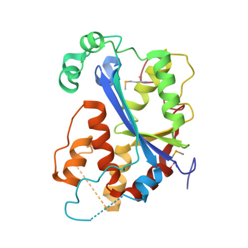
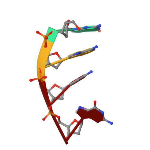
The Crystal Structure of TREX1 Explains the 3' Nucleotide Specificity and Reveals a Polyproline II Helix for Protein Partnering.

de Silva, U.Choudhury, S.Bailey, S.L.Harvey, S.Perrino, F.W.Hollis, T.

(2007) J Biological Chem 282: 10537-10543

  • DOI: https://doi.org/10.1074/jbc.M700039200
  • Primary Citation Related Structures: 
    2IOC, 2OA8

  • PubMed Abstract: 

    The TREX1 enzyme processes DNA ends as the major 3' --> 5' exonuclease activity in human cells. Mutations in the TREX1 gene are an underlying cause of the neurological brain disease Aicardi-Goutières syndrome implicating TREX1 dysfunction in an aberrant immune response. TREX1 action during apoptosis likely prevents autoimmune reaction to DNA that would otherwise persist. To understand the impact of TREX1 mutations identified in patients with Aicardi-Goutières syndrome on structure and activity we determined the x-ray crystal structure of the dimeric mouse TREX1 protein in substrate and product complexes containing single-stranded DNA and deoxyadenosine monophosphate, respectively. The structures show the specific interactions between the bound nucleotides and the residues lining the binding pocket of the 3' terminal nucleotide within the enzyme active site that account for specificity, and provide the molecular basis for understanding mutations that lead to disease. Three mutant forms of TREX1 protein identified in patients with Aicardi-Goutières syndrome were prepared and the measured activities show that these specific mutations reduce enzyme activity by 4-35,000-fold. The structure also reveals an 8-amino acid polyproline II helix within the TREX1 enzyme that suggests a mechanism for interactions of this exonuclease with other protein complexes.


  • Organizational Affiliation
    • Department of Biochemistry, Center for Structural Biology, Wake Forest University Health Sciences, Winston-Salem, North Carolina 27157.

Macromolecule Content 

  • Total Structure Weight: 53.76 kDa 
  • Atom Count: 3,891 
  • Modeled Residue Count: 454 
  • Deposited Residue Count: 474 
  • Unique protein chains: 1
  • Unique nucleic acid chains: 1

Macromolecules


Find similar proteins by:|  3D Structure
Entity ID: 2
MoleculeChains  Sequence LengthOrganismDetailsImage
Three prime repair exonuclease 1C [auth A],
D [auth B]
233Mus musculusMutation(s): 2 
Gene Names: Trex1
EC: 3.1.11.2
UniProt & NIH Common Fund Data Resources
Find proteins for Q91XB0 (Mus musculus)
Explore Q91XB0 
Go to UniProtKB:  Q91XB0
IMPC:  MGI:1328317
Entity Groups
Sequence Clusters30% Identity50% Identity70% Identity90% Identity95% Identity100% Identity
UniProt GroupQ91XB0
Sequence Annotations
Expand
Reference Sequence
Find similar nucleic acids by:  Sequence
Entity ID: 1
MoleculeChains LengthOrganismImage
5'-D(*GP*AP*CP*G)-3'A [auth C],
B [auth D]
4N/A
Sequence Annotations
Expand
Reference Sequence

Experimental Data & Validation

Experimental Data

  • Method: X-RAY DIFFRACTION
  • Resolution: 2.10 Å
  • R-Value Free:  0.259 (Depositor), 0.260 (DCC) 
  • R-Value Work:  0.192 (Depositor), 0.203 (DCC) 
  • R-Value Observed: 0.195 (Depositor) 
Space Group: P 1 21 1
Unit Cell:
Length ( Å )Angle ( ˚ )
a = 64.85α = 90
b = 57.141β = 107.47
c = 68.47γ = 90
Software Package:
Software NamePurpose
REFMACrefinement
CrystalCleardata collection
d*TREKdata reduction
d*TREKdata scaling
PHASERphasing

Structure Validation

View Full Validation Report


Entry History 

Deposition Data

Revision History  (Full details and data files)

  • Version 1.0: 2007-02-20
    Type: Initial release
  • Version 1.1: 2008-05-01
    Changes: Version format compliance
  • Version 1.2: 2011-07-13
    Changes: Advisory, Refinement description, Version format compliance
  • Version 1.3: 2023-08-30
    Changes: Data collection, Database references, Derived calculations, Refinement description
  • Version 1.4: 2023-11-15
    Changes: Data collection
  • Version 1.5: 2024-11-20
    Changes: Structure summary