Structural Analysis of Semi-specific Oligosaccharide Recognition by a Cellulose-binding Protein of Thermotoga maritima Reveals Adaptations for Functional Diversification of the Oligopeptide Periplasmic Binding Protein Fold.
Cuneo, M.J., Beese, L.S., Hellinga, H.W.(2009) J Biological Chem 284: 33217-33223
- PubMed: 19801540 Search on PubMedSearch on PubMed Central
- DOI: https://doi.org/10.1074/jbc.M109.041624
- Primary Citation Related Structures:
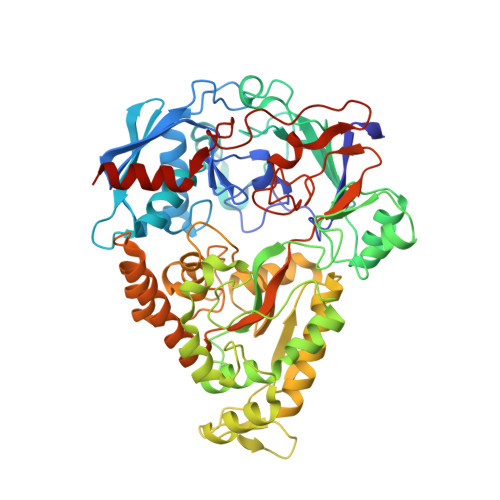
2O7I, 3I5O - PubMed Abstract:
Periplasmic binding proteins (PBPs) constitute a protein superfamily that binds a wide variety of ligands. In prokaryotes, PBPs function as receptors for ATP-binding cassette or tripartite ATP-independent transporters and chemotaxis systems. In many instances, PBPs bind their cognate ligands with exquisite specificity, distinguishing, for example, between sugar epimers or structurally similar anions. By contrast, oligopeptide-binding proteins bind their ligands through interactions with the peptide backbone but do not distinguish between different side chains. The extremophile Thermotoga maritima possesses a remarkable array of carbohydrate-processing metabolic systems, including the hydrolysis of cellulosic polymers. Here, we present the crystal structure of a T. maritima cellobiose-binding protein (tm0031) that is homologous to oligopeptide-binding proteins. T. maritima cellobiose-binding protein binds a variety of lengths of beta(1-->4)-linked glucose oligomers, ranging from two rings (cellobiose) to five (cellopentaose). The structure reveals that binding is semi-specific. The disaccharide at the nonreducing end binds specifically; the other rings are located in a large solvent-filled groove, where the reducing end makes several contacts with the protein, thereby imposing an upper limit of the oligosaccharides that are recognized. Semi-specific recognition, in which a molecular class rather than individual species is selected, provides an efficient solution for the uptake of complex mixtures.
- Department of Biochemistry, Duke University Medical Center, Durham, North Carolina 27710, USA.
Organizational Affiliation: